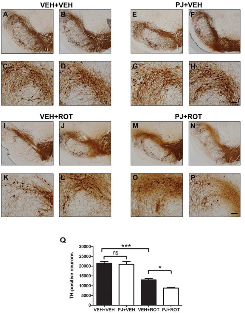
Figure 5.
Immunostaining of tyrosine hydroxylase-positive neurons in the substantia nigra. Examination at low (scale bar = 500 µm) and high magnification (scale bar = 50 µm) of the dorsolateral nigra revealed a dense TH-immunopositive network of cell bodies and fibers in the SN in controls alone (A-D) or with PJ (E-H). After the rotenone injections, there was a reduction in TH immunoreactivity at the level of the SN (I-L). Oral administration of PJ with rotenone caused an increase in DA cell loss and pruning of processes compared to treatment with rotenone alone (M-P). The loss of SN neurons was counted by unbiased stereology, and rats treated with PJ showed potentiation of rotenone-induced neurotoxicity (Q). Bars represent means ± S.E.M. Statistical analyses were carried out using the Newman Keuls post-hoc test. *** p < 0.001 vs VEH+VEH; * p < 0.05 vs VEH+ROT.

