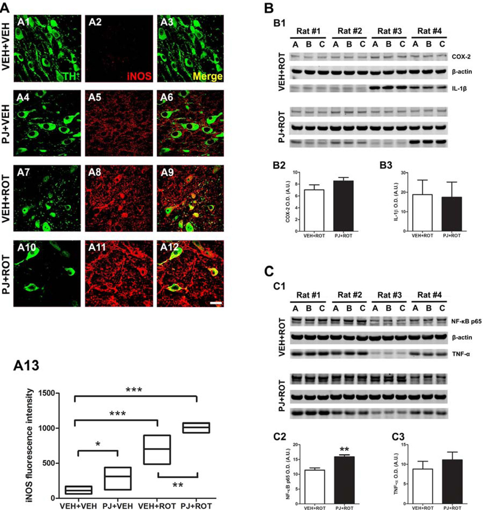
Figure 8.
Nigral proinflammatory mediators. Confocal images at 100x revealed an absence of iNOS expression in the vehicle group (A2) but oral administration of PJ significantly increased iNOS immunoreactivity (A5). Following rotenone injection, a marked expression of iNOS was detected (A8) and this effect was even greater when PJ was co-administered with rotenone (A11). Quantification of fluorescence levels of iNOS was expressed as the fold increased versus untreated controls (A13) and is representative of the average of 200–300 DA neurons corresponding to 5 SN sections per animal. Results are expressed as the mean ± S.E.M. of 4 rats per group. Scale bar = 20 µm. *** p < 0.001, * p < 0.05 vs VEH+VEH, Newman-Keuls posthoc test; ** p < 0.01 vs VEH+ROT. Representative western blots of different proinflammatory markers in SN brain homogenates (B and C). Densitometric analysis disclosed that the amount of protein for the enzyme COX-2 (~ 74 kDa), the cytokine IL-1β (~ 17 kDa), and TNF-α (~ 17 kDa) remained unchanged (B2, B3, and C3, respectively). Nevertheless, NF-kB p65 (~ 65 kDa) expression levels were significantly enhanced after dietary supplementation with PJ (C2). The histogram values represent the mean ± S.E.M in arbitrary units of the optical density of 4 independent measurements in triplicate (A-C are biological replicates). β-Actin was used as a reference control. Significant differences between groups were determined by one-tailed Student’s t-test. ** p < 0.01 and * p < 0.05 compared to rotenone.

