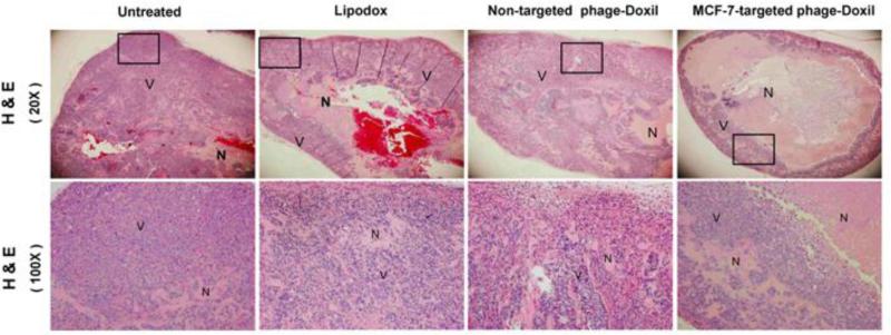
Figure 3. The confirmation of antitumor activity by H & E staining.
Representative images of tumor sections. Necrotic cells (N) showing eosinophillic cytosol (pink) accompanied by the absence of hemotoxylin-stained nuclei (blue); viable cells (V) showing eosinophillic cytosol (pink) accompanied by hemotoxylin-stained nuclei (blue).

