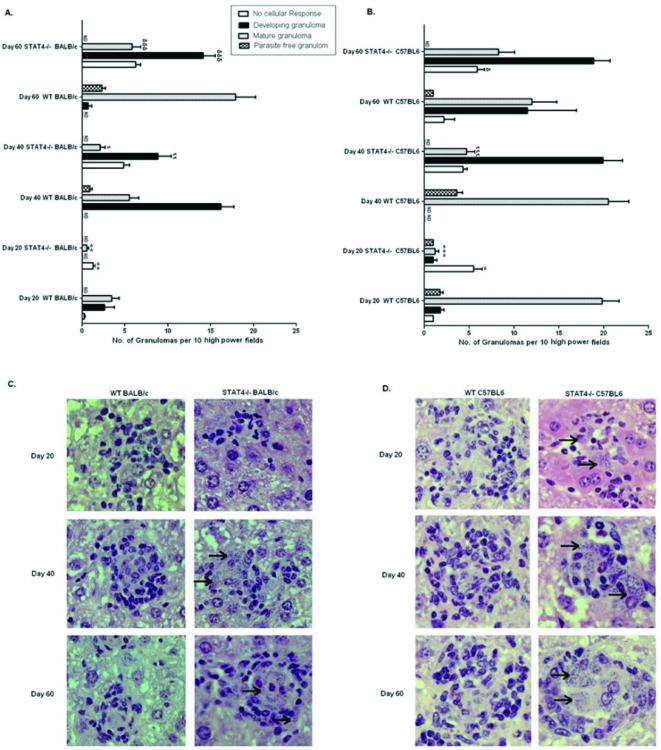
Figure 4. Immunopathology of stat4−/− mice during L. donovani infection.
Liver sections from L. donovani infected WT and stat4−/− BALB/c mice (A) or WT and stat4−/− C57BL/6 mice (B) at days 20, 40 and 60 post infection were scored for the extent of granuloma formation. Results were from one representative experiment of three with similar findings. Data were expressed as mean granuloma number per 10 high-power fields (magnification 200×) ±SE. ND, not determined.*, P < 0.05, **, P < 0.01 and ***, P < 0.001 compared to their respective infected Day 20 WT control; ι, P < 0.05, ιι, P < 0.01 and ιιι, P < 0.001 compared to their respective infected Day 40 WT control; φ, P < 0.05, φφ, P < 0.01 and φφφ, P < 0.001 compared to their respective infected Day 60 WT control.
Histopathology of infected livers from WT and stat4−/− BALB/c mice (C), or WT and stat4−/− C57BL/6 mice (D). WT BALB/c and C57BL/6 mice showed developing granuloma at day 20 post infection and contained matured and parasite free granulomas at days 40 and 60 post infection. In contrast, stat4−/− BALB/c and C57BL/6 mice showed marked delay in granuloma formation as there was lack of cellular reaction at day 20 post infection and even at days 40 and 60 post infection, the granulomas were poorly developed and were highly parasitized (arrow). Magnifications 400× for panels C and D.

