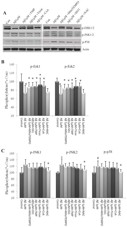
Figure 5. Short-term exposure of NPCs to Aβ1–42 OCP inhibits ERK signaling.
Dissociated adherent NPCs were pretreated for 1 h with 1 μM MitoTEMPO, 50 μM SS31, 1 μM TMP, 100 μM Tiron, 0.1μM CsA or 1μM NAC, and were then treated for 24 h with 1μM Aβ1-42 oligomers. Cell lysates were then prepared and subjected to immunoblot anlaysis using antibodies that selectively recognize phosphorylated (active) forms of ERKs 1 and 2, p38 or JNK. Blots were reprobed with phosphorylation-insensitive antibodies against actin. (A – C) Representative immunoblot (A) and densitometric analysis (B and C) of NPCs. #p<0.05 compared to the mean value of control NPCs without Aβ1-42 OCP treatment; *p<0.05 compared to the mean value of NPCs treated with Aβ1-42 OCP. n= 3 separate experiments.

