
Figure 3. Apoptosis is reduced by H2S treatment.
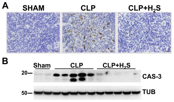
A) Activated Caspase-3 immunoperoxidase staining was performed on spleen sections 18h after CLP (40 X objective, bar 20 mm). B) Lysates from spleens 18 h after CLP were subjected to Western blotting assay to detect activated Caspase-3 (CAS-3). Tubulin (TUB) was used as a loading control. Samples represent individual mouse spleen from 2 sham, 5 CLP and 5 CLP+H2S.