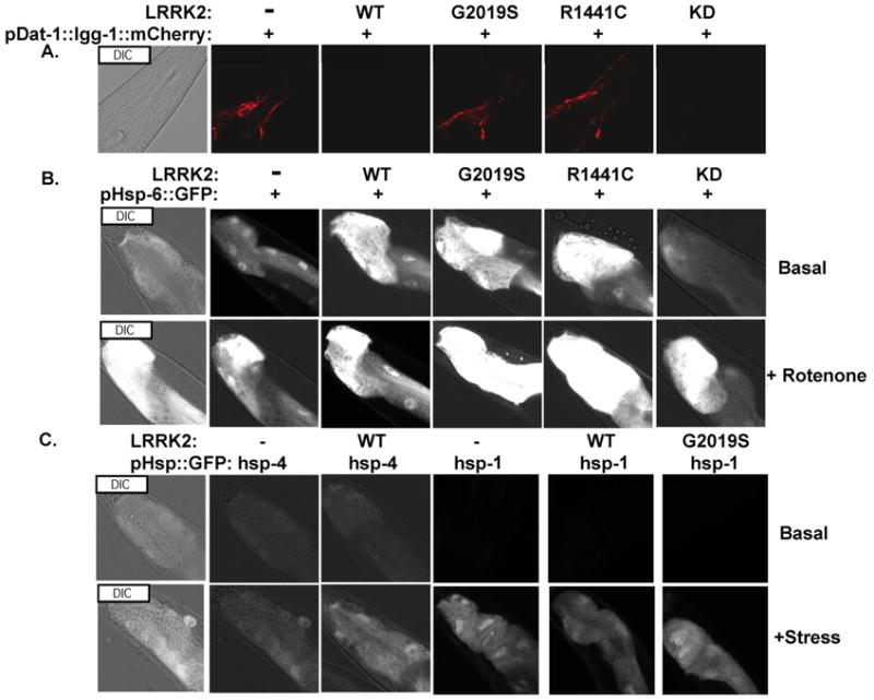
Fig. 1.

Chromosomally integrated reporters, which reflect autophagic activity in in the CEP dopaminergic neurons and the stress responses elicited in mitochondria, ER and cytoplasm (b, c) with the Distal Tip Cells (DTC) in the posterior part of the nematode a) The pDAT::lgg-1::mCherry reporter: Autophagic fluorescence from day 5 adults. Autophagic puncta are seen in the cell body of CEP neurons and nerve ring in the nematode's head region. WT and KD (kinase dead) LRRK2 reduce mCherry fluorescence, reflecting increased autophagic flux. (b) The pHSP6::GFP reporter: Mitochondrial HSP 70 response in DTC cell with basal and induced (250nM Rotenone, 24 hrs) conditions. WT, G2019S and R1441C LRRK2 increase basal activity. (c) The pHSP4::GFP and pHSP1::GFP reporters: stress responses from ER compartment (hsp-4) and cytoplasmic compartment (hsp-70) from DTC cells with basal and induced conditions with 2.5 mg/ml Tunicamycin treatment and heat shocking at 330C respectively. LRRK2 constructs did not affect fluorescence of these reporters.
