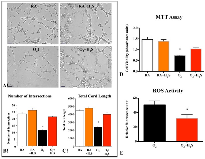
Figure 1. H2S protected human pulmonary artery endothelial cells (HPAECs) from O2-induced toxicity.
(A) H2S promotes endothelial network formation. Quantitative assessment of cordlike structure formation shows a significant decrease in the number of intersects and the total length of cord-like structures in hyperoxia. H2S preserved the number of intersects (B) and total cord-structure length (C). (n = 3 per group, *P<0.0001 hyperoxia vs. other groups, scale bar 65 µm). (D) HPAECs were cultured for 48 hours in room air (Normoxia) or 95% hyperoxia. Mean data of cell viability as assessed by measuring the mitochondrial-dependent reduction of colorless 3-(4,5-dimethylthiazol-2-yl)-2,5-diphenyltetrazolium bromide (MTT) shows that hyperoxia significantly decreases HPAECs viability as compared with room air–exposed cells. H2S treatment significantly improved HPAECs viability in hyperoxia (n = 7, *P<0.001). (E) After 48 hours culture in hyperoxia (95%), ROS activity evaluated by measuring the dichlorofluorescein (DCF) shows that hyperoxia increases the ROS production in HPAECs, treatment with GYY4137 significantly decreased the ROS (n = 6/group, *P<0.005 Hyperoxia vs O2+H2S).

