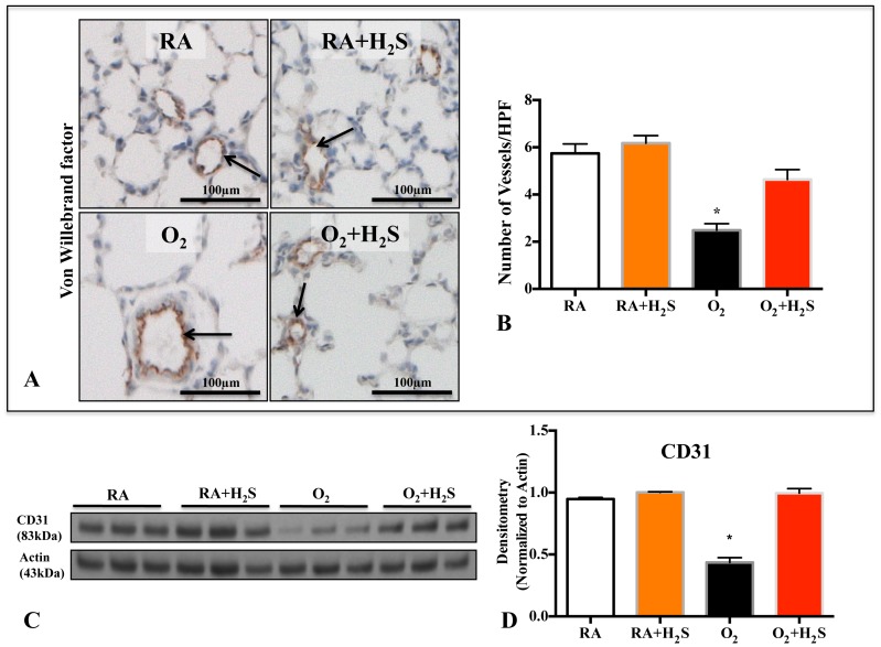
Figure 3. In vivo H2S treatment prevents O2-induced arrested lung vascular growth.
A. Representative photomicrographs showing von Willebrand (vWF) factor staining (brown) in RA (room air), RA+H2S, hyperoxia (O2) and O2+H2S exposed lungs. Arrows highlight vWF-positive vessels; scale bars represent 100 µm. B. Mean data quantifying the number of vWF positive vessels between groups. The decrease in the number of vessels per high-power field (HPF) after hyperoxia exposure was prevented by H2S treatment (n = 5–7/group, *P<0.005 Hyperoxia vs O2+H2S). C. Representative immunoblot and densitometric (D) analysis for endothelial marker CD31 in lung homogenates from control and H2S treated animals. H2S treatment preserved the expression of CD31 in hyperoxic rats compared with hyperoxic control (n = 3/group, *P<0.005 Hyperoxia vs O2+H2S).

