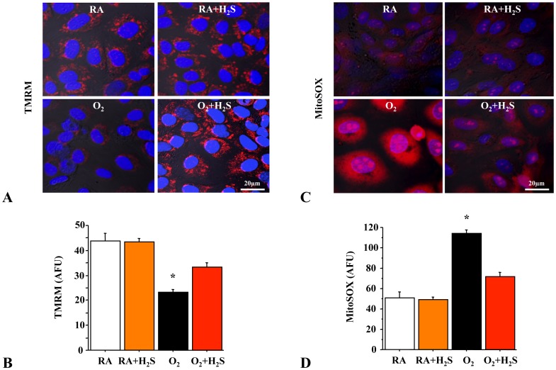
Figure 7. Rat lung epithelial cells (RLEs) exposed to hyperoxia decreased mitochondrial ΔΨm and increased mROS.
Representative confocal microscopy at high magnification (×100) of rat lung epithelial cells (RLEs) showing (A) decreased ΔΨm (TMRM) and (C) increased mROS production (MitoSOX) in hyperoxia (TMRM and MitoSOX are in red, merged with nuclear stain DAPI in blue). Hyperoxia exposed RLEs treated with H2S have significantly increased ΔΨm (B) and decreased mROS (D) compared to hyperoxic control (n = 4 per group, *P<0.005 hyperoxia vs. other groups).

