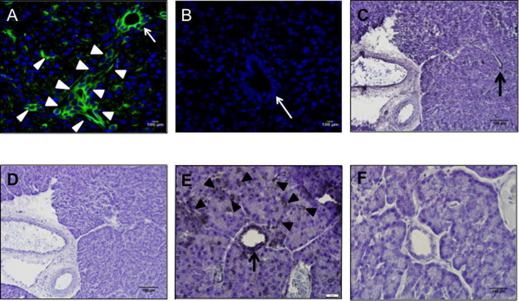
Figure 3. AAV9 transduces porcine ductal epithelial cells.

Pancreas sections 30 days after newborn pigs received AAV9CMV.sceGFP (A, C, D, E, F) (2.4×10¹² vg per animal) or vehicle (B) into the celiac artery. Immunofluorescence (A, B) and immunohistochemistry (C–F) images are shown. Arrows point to intralobular (larger) ducts, arrowheads point to intercalated (smaller) ducts. C and D; E and F are serial sections from the same animal, primary antibody is omitted in D and F. A, B ×20 mag; C, D ×10 mag, scale bar = 100 µm; E, F ×60 mag, scale bar = 20 µm. Green: GFP, Blue: DAPI nuclei.
