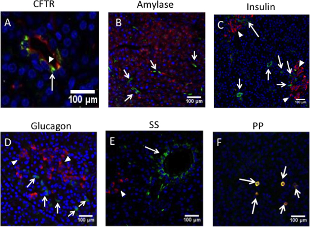
Fig. 6. AAV9 vector expression of GFP in CFTR-expressing duct cells.

Immunofluorescent images of pancreas from pigs, 30 days after receiving 2.4×10¹² vg AAV9CMV.sceGFP in the newborn period. (A) anti-CFTR antibody (red) for pancreatic ducts; (B) anti-amylase (red, arrowheads) for acinar cells; (C) anti-insulin (red, arrowheads) for β cells; (D) anti-glucagon (red, arrowheads) for α cells; (E) anti-somatostatin (SS) (red, arrowheads); (F) anti-pancreatic polypeptide (PP) (red-yellow indicating colocalization with eGFP, arrows); DAPI (blue) for nuclei. AAV9-GFP (green, arrows) was transduced in the cells that were expressing CFTR (red, arrowhead) on the apical side, A ×40 mag; B, C, D, E, F= ×20 mag. A, B, C, D, E= cells expressing GFP are shown with arrows.
