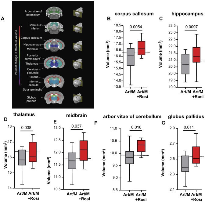
Figure 6. Rosiglitazone adjunctive therapy protects mice from malaria-induced brain atrophy.
Following completion of all behavioural testing mouse brains were scanned using magnetic resonance imaging (MRI). Image registration and volumetric analysis of the brain volume of 62 distinct regions were performed. Linear regression analysis was used to identify areas that differed significantly between the mice treated with artesunate/mefloquine plus saline (grey bars), and the mice treated with artesunate/mefloquine plus rosiglitazone (red bars). (A) Identified brain regions are shown. Percentage change in brain structure volume is indicated by colour (see rainbow bar on the left). Significant differences were observed in (B) the corpus callosum, (C) the hippocampus, (D) the thalamus, (E) the midbrain, (F) the arbour vitae of the cerebellum, and (G) the globus pallidus of the basal ganglia. N = 13 for artesunate/mefloquine, N = 10 for artesunate/mefloquine + rosiglitazone. The median volume for uninfected mice is shown as a dashed line (N = 10 for uninfected control). Additional brain regions are shown in Figure S8.

