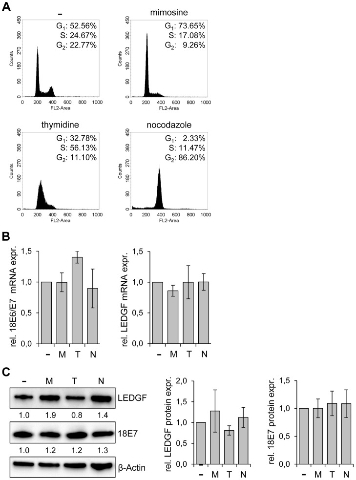
Figure 4. LEDGF expression in HeLa is not altered by cell cycle-inhibitory drugs.
(A) Cell cycle distribution analyzed by FACS. HeLa cells were either left untreated (-) or treated with mimosine, thymidine or nocodazole. The percentages of cells in the G1, S, and G2 phases are indicated. (B) qRT-PCR analyses of E6/E7 (left panel) and LEDGF (right panel) transcript levels in untreated cells (-) and in cells treated with either mimosine (M), thymidine (T) or nocodazole (N). Indicated are relative mRNA levels above those of untreated cells, arbitrarily set at 1.0. Standard deviations are indicated. Statistical analyses did not reveal significant differences between untreated and treated cells. (C) Analysis of LEDGF and HPV18 E7 protein levels upon treatment of HeLa cells with either mimosine (M), thymidine (T) or nocodazole (N). (-): untreated cells. β-Actin: loading control. Statistical analyses did not reveal significant differences between untreated and treated cells. Left panel: Representative immunoblot. Relative quantifications of LEDGF and HPV18 E7 signal intensities are indicated below the respective lanes, the value for untreated cells was set at 1.0. Right panel: Statistical analyses from three different immunoblots did not reveal significant differences between untreated and treated cells.

