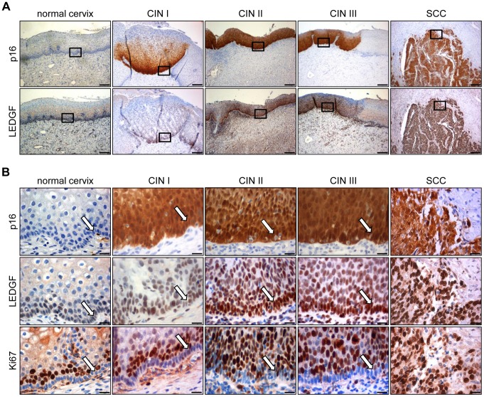
Figure 8. Immunohistochemical analysis of LEDGF expression.
(A) Expression of LEDGF in histologically normal cervical epithelium, in dysplastic CIN I to CIN III lesions, and in cervical squamous cell carcinoma (SCC). p16: surrogate marker for HPV oncogene expression. Bars correspond to 200 µm. (B) Higher magnification of normal cervix, CIN I to III lesions, and cervical SCC. Staining of p16, LEDGF and proliferation marker Ki67. Arrows indicate the basal cell layer in normal cervix and CIN lesions. Bars correspond to 20 µm.

