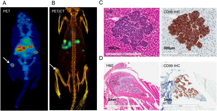
Figure 4. 64Cu-DN16 PET imaging of micrometastatic lesions.
A-B. An independent mouse with 1-2 mm liver metastases was imaged 24 hrs after injection of 64Cu-DN16. An ectopic focus of activity in the femur was noted (white arrow). C. Hematoxylin-eosin (H&E) staining of a metastatic liver lesion, as well as anti-CD99 immunohistochemistry (CD99 IHC). D. H&E and CD99 IHC right femur, corresponding to the PET-focus in A-B.

