
Figure 1. Sagittal (a) and axial-oblique (b) T2-weighted images showing dimensions of a large fibroid.
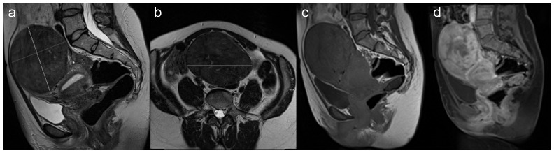
(c) Magnetization transfer-weighted and (d) dynamic contrast-enhanced MRI (DCE-MRI) image (acquired one minute after injection of contrast) of the same patient.