
Figure 4.
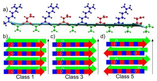
a) A model of a RADA16-I molecule in a β-strand conformation, with all atoms drawn for the arginine (blue), alanine (green), and aspartate (red) sidechains. This conformation creates hydrophobic (green) and hydrophilic (alternating blue (+) and red (−)) faces represented schematically in b-d. b-d) Schematic representations of nanofiber symmetry classes, originally defined by Sawaya et al., for RADA16-I β-strands arranged into stacks of 2 β-sheets. The white circles represent positions of A4 Cβ sites. Black arrows represent the long axis of the nanofiber.