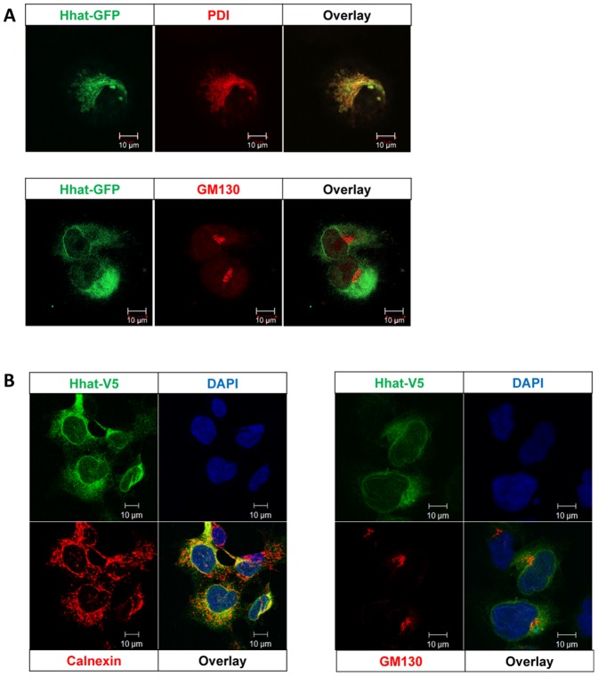
Figure 1. Localization of Hhat in PANC1 cells.
A. Localization of Hhat in PANC1 cells was assessed using Hhat-EGFP transfection and confocal microscopy combined with immunofluorescence localization of ER (PDI) and Golgi (GM130). B. HEK293a Hhat-V5 stable cells were co-stained for the V5 epitope with ER (Calnexin) or Golgi (GM130) and nuclei (DAPI). The data show that both Hhat-EGFP and Hhat-V5 localize primarily in ER with little if any in Golgi apparatus. Scale bar = 10 µm.

