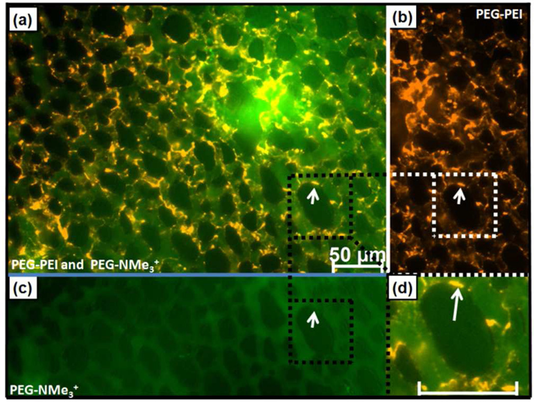
Figure 4.
Differential in vivo binding and flow of size- and charge-matched PEG-PEI (orange) and PEG-NMe3+ (green) MSNP in chick CAM 10 min post injection. (a) Merged image (b) PEG-PEI showing arrest on endothelial cells, and (c) PEG-NMe3+ image showing circulating MSNPs. (d) Magnification of PEG–PEI MSNP binding (arrow) on endothelial cells (scale bar – 50 µm).

