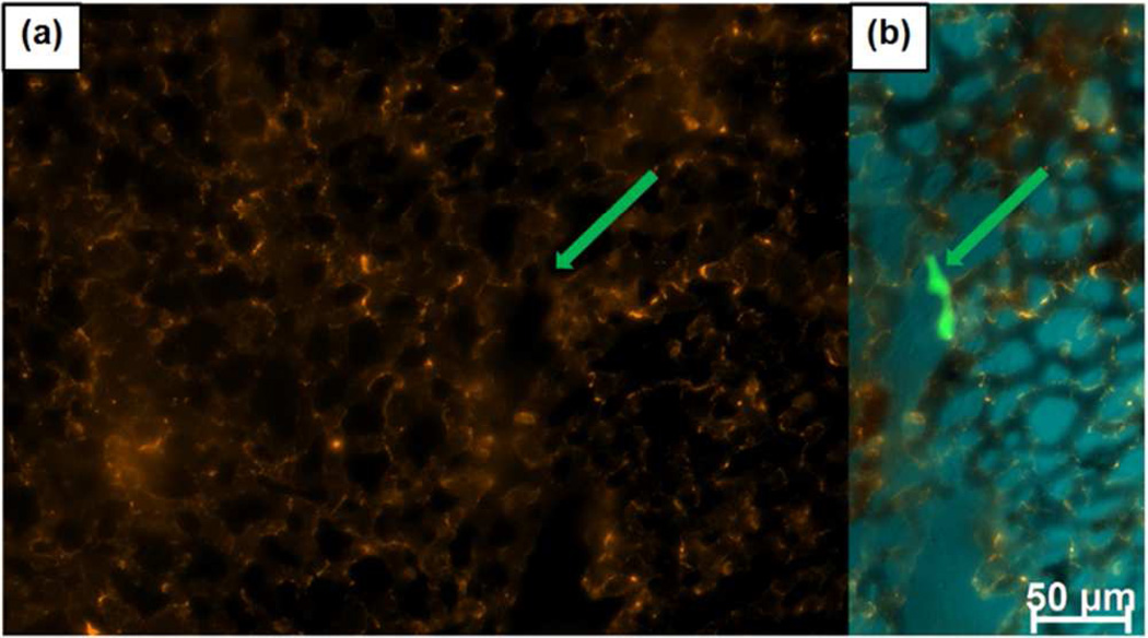
Figure 5.
PEG-PEI MSNPs deposit on the vessel walls and are scavenged by white blood cells of CAM immediately following injection, with no apparent binding to cancer cells. (a) Particles binding to vasculature and WBCs with no specific out-line of a cancer cell (location indicated by arrow). (b) Merged particle and cancer cells (green) image. CAM tissue autofluorescence is false colored blue.

