
Figure 3.
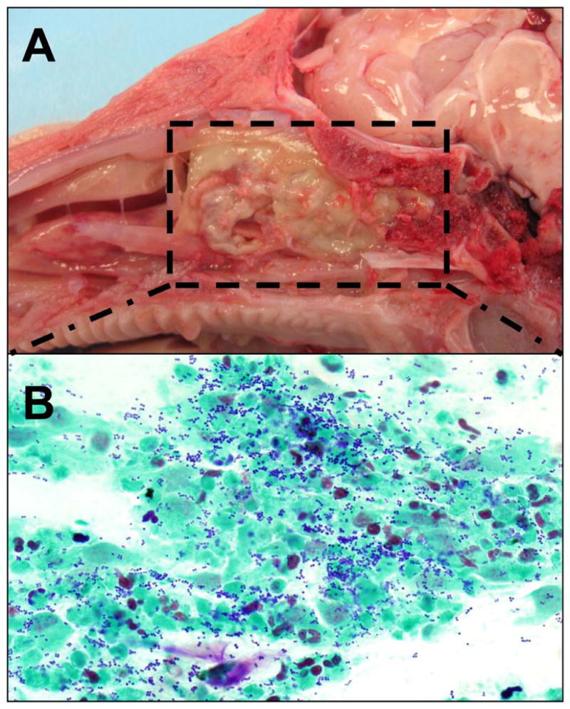
CF piglet with thick mucus occluding the ethmoid and maxillary sinus (A). Histology of the mucus consistent with cellular debris and gram positive cocci consistent with Staph aureus (B).
Official websites use .gov
A
.gov website belongs to an official
government organization in the United States.
Secure .gov websites use HTTPS
A lock (
) or https:// means you've safely
connected to the .gov website. Share sensitive
information only on official, secure websites.
CF piglet with thick mucus occluding the ethmoid and maxillary sinus (A). Histology of the mucus consistent with cellular debris and gram positive cocci consistent with Staph aureus (B).