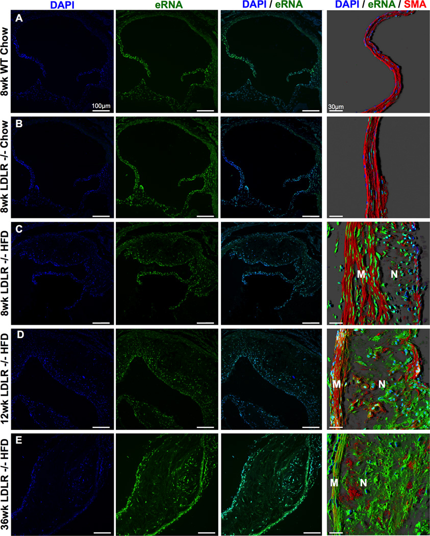
Figure 1.
Time-progressive distribution of extracellular RNA (eRNA) in atherosclerotic lesions. The presence of eRNA in aortic root tissue from WT or Ldlr−/− mice fed a chow diet for 8 weeks or from Ldlr−/− mice fed a high-fat-diet (HFD) for 8 (A-C), 12 (D) or 36 weeks (E), as indicated, was demonstrated by confocal microscopy using an RNA-binding fluorescence dye (RNA-Select, green) together with cell nuclei staining (DAPI, blue). Right panel in each line: confocal images with merged immunostaining for eRNA (green), cell nuclei (DAPI, blue) and smooth muscle cell actin (SMA, red); M: media; N: neointima. (n=6 per group).

