Figure 2.
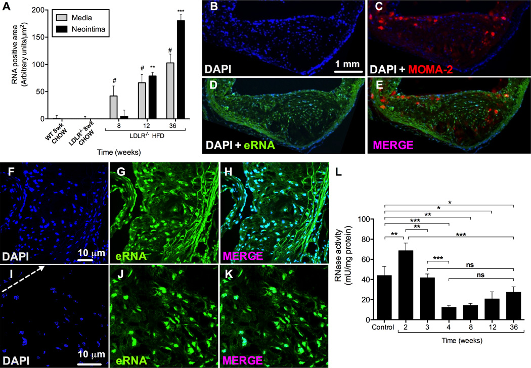
Co-localization of eRNA and macrophages in atherosclerotic lesions of Ldr−/− mice after 36 weeks on HFD, and RNase activity in mouse plasma. (A) Quantitative analysis of eRNA-associated fluorescence intensity in aortic root tissue in the media and neointima in wild-type (WT) and Lldr−/− mice fed a normal chow or high fat diet for indicated time periods. Values are expressed as mean ± SD (n=6 per group); #p<0.05 vs. media, and **p<0.001 vs. neointima in chow-fed Lldr−/− mice. (B-E) Imunofluorescence staining in cryosections of atherosclerotic lesions from Ldlr−/− mice after 36 weeks on HFD. Staining for eRNA (D) was performed by an RNA-binding fluorescence dye (RNA-Select, green), macrophages (C) were identified by the anti-MOMA-2 monoclonal antibody (red), and cell nuclei (B) were marked by DAPI staining (blue). (E-K) Higher magnifications of atherosclerotic lesions are shown stained with DAPI (blue, F,I; arrow indicates cellular towards acellular necrotic core regions) and RNA-binding fluorescence dye (green, G,J); merged images are indicated in (H,K). All images were obtained under identical conditions of confocal laser beam intensity and exposure time; representative images are displayed (n=4). (L) RNase activity in Ldlr−/− mouse plasma was quantified for each time point and normalized to plasma protein concentration. Values are expressed as mean ± SD (n=6–12 per group); ns=non-significant, *p<0.05, **p<0.01, ***p<0.001.

