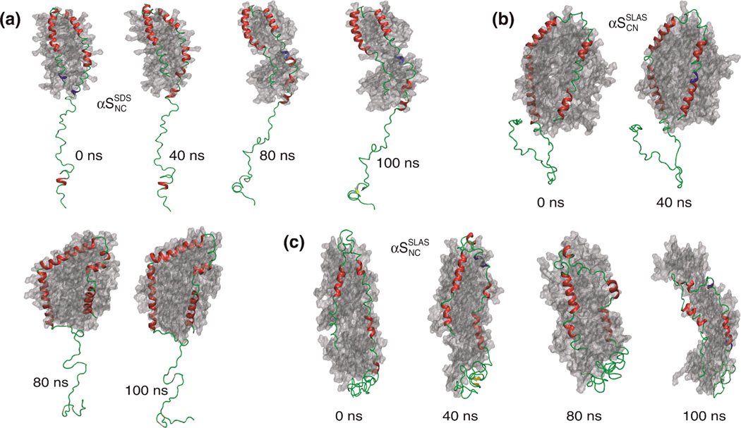
Figure 6.
Snapshots from all-atom MD simulations of micelle-bound αS. Snapshots for (a) αSSDSNC, (b) αSSLASNC and (c) αSSLASCN during the simulation (0–100 ns). The micelle surface is shown in grey, α-helix in red, 310-helix in blue, β-sheet in yellow, and turn and coil in green. Protein secondary structures were classified with the program STRIDE.70

