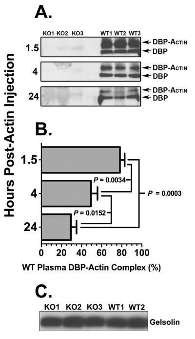
Figure 1.
Plasma DBP-actin complexes and gelsolin levels in DBP+/+ and DBP−/− mice following intravenous actin injection. (A) DBP immunoblot of EDTA plasma samples from three DBP+/+ wild-type (WT) mice and three DBP−/− (KO) mice each obtained 1.5, 4 and 24 hours after actin injection. Plasma aliquots were separated using a 10% native (non-denaturing) gel then blotted for DBP. The electrophoretic separation of unbound DBP and DBP-actin complexes is shown. (B) Densitometry measurement of DBP-actin complexes in panel A. Data is presented as mean ± SEM (n = 3) of DBP-actin as a percent of total DBP (unbound + DBP-actin) in WT plasma. Statistical significance is indicated. (C) Gelsolin immunoblot of plasma separated by SDS-PAGE from three DBP−/− (KO) and two DBP+/+ (WT) mice.

