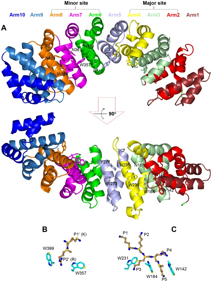
Figure 1. Structure of an importin α-NLS complex (PDB entry 1Q1T).
(A) The 10 Arm repeats of the importin α NLS-binding domain, shown in different colors. The conversed W-N residues are shown as sticks. (B) and (C): the highly conserved importin α-NLS interactions at the minor and major sites, respectively. Importin α residues are shown as sticks with carbon atoms in cyan, and NLS residues are shown as ball-and-stick with carbons atoms in sand. Carbon atoms used for calculating positional dispersions at the minor site in 24 crystal structures and at the major site in 35 crystal structures are shown in yellow. All structure figures were generated by Pymol (http://www.pymol.org/).

