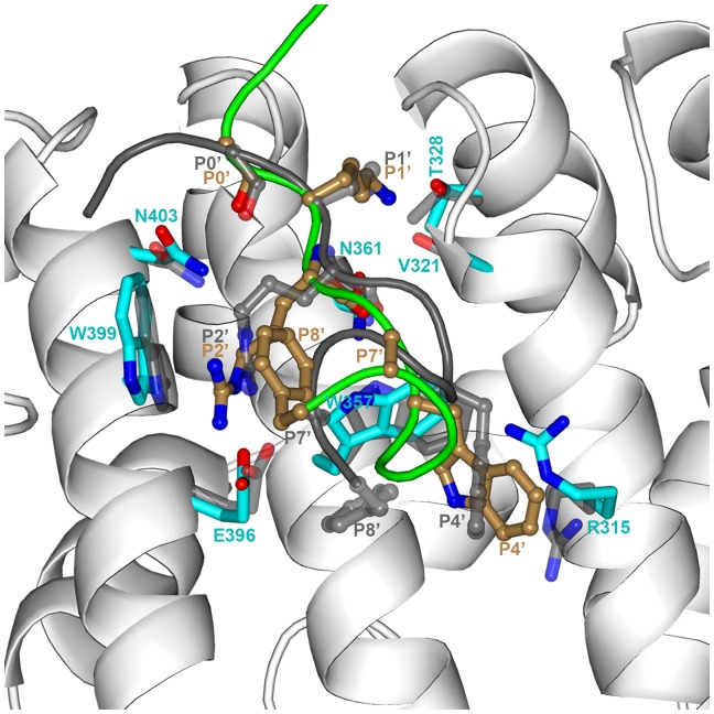
Figure 5. Comparison the unrestrained MD snapshot at 20-ray structure for the minor-site bound NLS3-importin α complex.
The MD simulation started from our modeled structure; superposition to the X-ray structure was done on the Cα atoms of importin α residues within 5 Å of NLS3. The color scheme for the MD snapshot is the same as in Figure 3. For the X-ray structure, the backbone of importin α is undisplayed for clarity and the backbone of NLS3 is shown as dark gray tube; key residues of the peptide and protein are shown as ball-and-stick and as sticks, respectively, both with carbon in gray.

