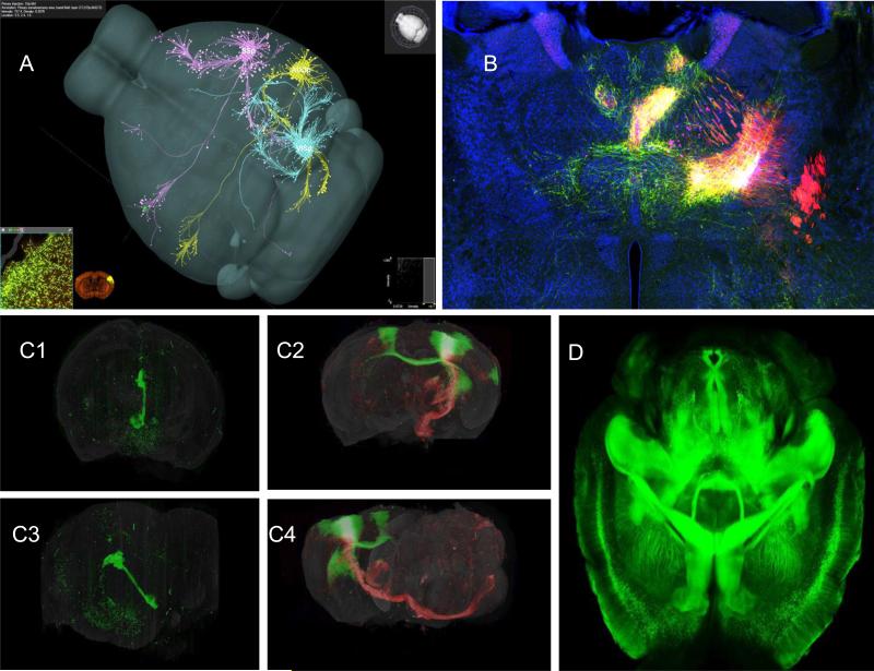
Fig 5. Mouse connectome.
A. Three AAV-GFP tracer injections into the mouse primary somatosensory (SSp), the primary auditory (AUDp) or the primary visual (VISp) cortical area, respectively, are co-displayed in a common 3D mouse brain model in the Brain Explorer program. Courtesy of Hongkui Zeng, Allen Brain Institute B. A representative digital image of the iConnectome project (www.MouseConnectome.org) showing multiple fluorescent labeled neuronal pathways through one coronal thalamic level of the mouse brain, after multiple fluorescent tracers were injected into the infralimbic (PHAL and CTb) and prelimbic (AAV and CTb) cortical areas. Green axons are labeled with PHAL; red axons are labeled with AAV; magenta neurons are labeled with CTb. The yellow regions also contain fluorogold labeled neurons, which are mostly intermixed with AAV axons. This approach effectively reveals two sets of afferents and two sets of efferent pathways associated with two anterograde/retrograde tracer coinjection sites within the same brain and simultaneously exposes reciprocal connections . Courtesy of Hongwei Dong, Univ. Southern California. C. The 3-D reconstruction of two whole mouse brains injected in the left hemisphere with fluorescent virus tracers to visualize specific sets of connectivity. Top, frontal view; bottom, left lateral view. (C1) Left, GFP labeled modified ‘retrograde’ rabies virus injected in the medial nucleus of the habenula (mH) showing retrogradely labeled cell bodies in the hypothalamus and frontal cortex, as well as the passively filled output projection from the mH (Fasciculus Retroflexus) . (2) Right, double injection of primary motor cortex (MOp) with modified ‘anterograde’ adeno-associated virus (AAV2/1 CAG) injection to highlight axons of efferent projections by supragranular neurons (injection with GFP-AAV), and infragranular neurons (injection with RFP-AAV). Courtesy of Partha Mitra, Cold Spring Harbor Laboratory. D. CLARITY is a newly developed technology to transform intact and opaque biological tissue into a hybrid form of formaldehyde-fixed tissue and hydrogel monomers which is physically stable and permeable to exogenous macromolecules. Because the hybrid is permeable to exogenous macromolecules, the whole tissue can be uniformly stained with antibodies to reveal specific cell types and allows multi-round molecular phenotyping of an intact brain. Adapted with permission from Chung and Deisseroth154 .

