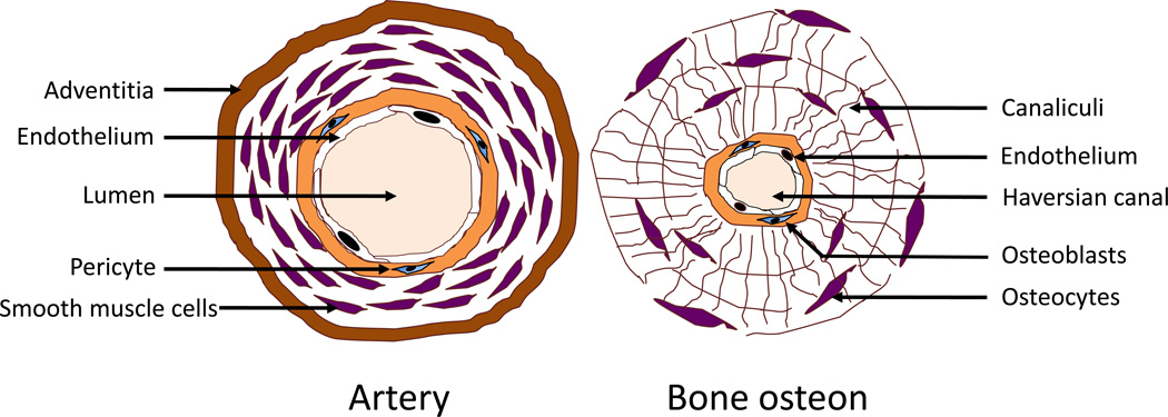
Figure 1. Schematic comparing the anatomic structure of an artery and bone osteon.
Both the artery and osteon are centered on a blood lumen, which is surrounded by a single layer of endothelium. This, in turn, is surrounded by a basement membrane housing immature mesenchymal cells. In arteries, the immature cells are pericytes and/or smooth muscle cells, whereas, in bone, they are pericytes and/or preosteoblasts in the bone. (Modified from Parhami et al., Arteriosclerosis, Thrombosis, and Vascular Biology, 1997; 17:680–687).

