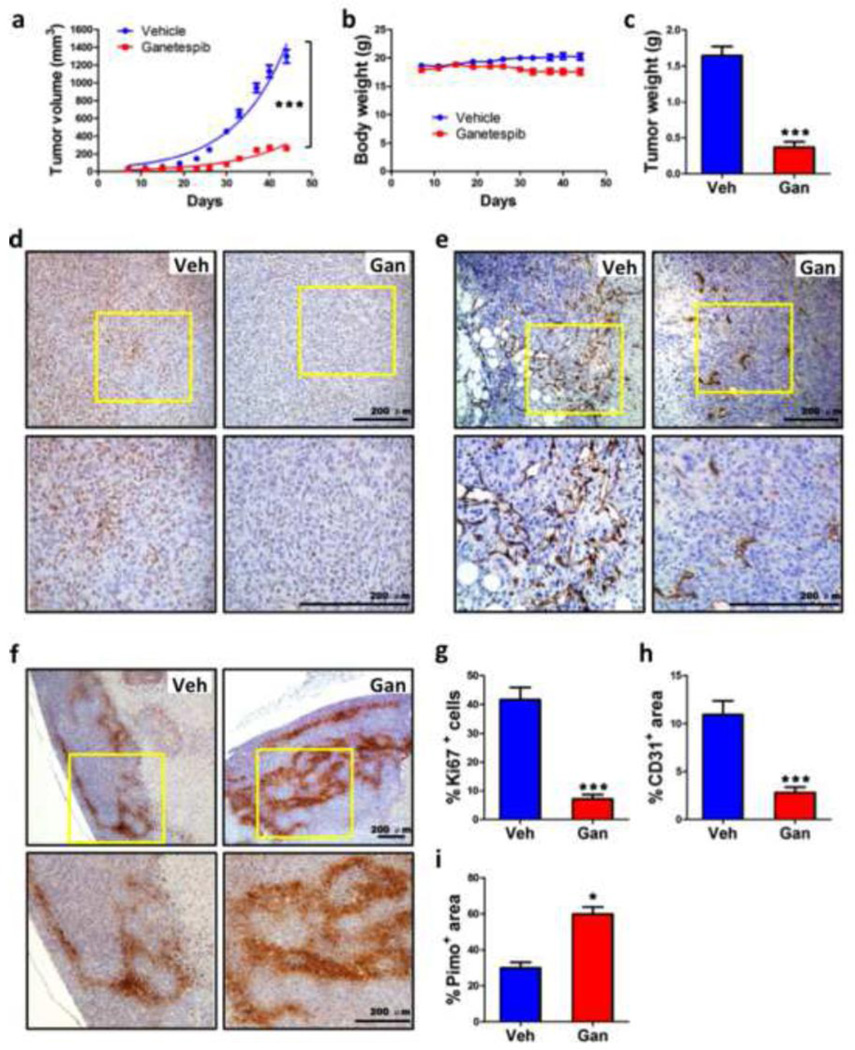
Fig. 2.
Ganetespib treatment inhibits breast tumor growth and vascularization. Severe combined immunodeficiency (SCID) mice received a mammary fat pad injection of 2×10⁶ MDA-MB-231 cells. When tumors became palpable, the mice were treated by tail vein injection of vehicle (Veh) or ganetespib (Gan; 150 mg/kg/week) starting on day 7 after orthotopic injection and weekly thereafter. a-c Tumor volume (a) and body weight (b) were determined twice weekly. On day 44, the mice were administered pimonidazole and, 1 h later, tumors were harvested and weighed (c). ***p < 0.001 vs vehicle, two-way ANOVA or Student’s t test (mean ± SEM; n = 9). d-f Immunohistochemistry was performed on tumor tissue sections from mice treated with Veh or Gan to analyze cell proliferation by Ki67 staining (d), angiogenesis by CD31 staining (e), and intratumoral hypoxia by pimonidazole staining (f). Representative photomicrographs are shown, with the bottom panel showing higher magnification of the insert from the top panel; scale bar = 200 µm. g-i The stained sections were subjected to image analysis and quantification (mean ± SEM); *p < 0.05, ***p < 0.001 vs vehicle, Student’s t test.

