
Figure 3. Nef is present in endothelial cells in vivo.
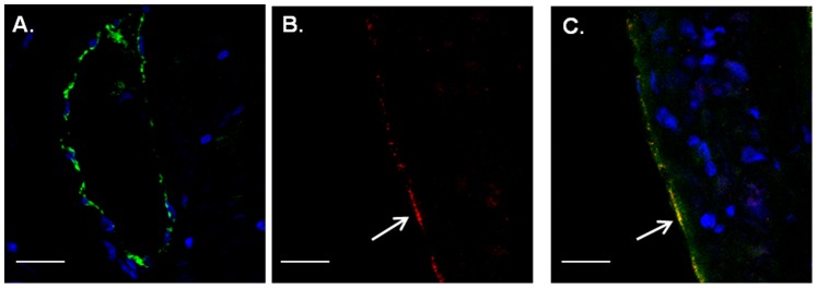
A–C. Heart sections of single CD4-GFP (A) and double CD4-Nef-GFP (B, C) transgenic mice (N = 3; at least 4 pictures/slide) were double stained with GFP antibody (green, white arrows) and the endothelial marker vWF (red). Shown is GFP within the endothelial lining (arrows). Original magnification, X 60. Scale bars represent 100 µm. D–F. Macaque heart sections (N = 5; at least 4 pictures/slide) were double stained with IgG control (D) or Nef (E, F, red) and the endothelial marker vWF (green). Shown are cells double positive for Nef and vWF in coronary arteries (arrow). Original magnification, X 60. Scale bars represent 100 µm.