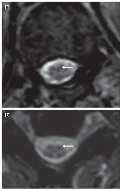
Fig. 2.

MRI after injection of Feridex®-labeled mesenchymal stem cells. a An axial T2-weighted gradient echo scan through the inferior thoracic cord shows a hypointense pial signal coating the cord similar to that of superficial siderosis, characteristic of Feridex®-labeled cells. b Axial T2-weighted gradient echo scan through the cervical cord shows hypointensity of the dorsal roots and their entry zone and a similar hypointensity of the ventral root entry zones, suggesting the presence of Feridex®-labeled cells. (Reproduced from Ref. 52, with permission)
