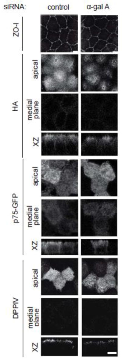
Figure 4. Steady-state distribution of raft-associated and raft-independent apical cargoes is not affected by α-gal A siRNA silencing. (A).

Indirect immunofluorescence staining of ZO-I (tight junction marker) in control and α-gal A silenced MDCK cells confirms that tight junctions are intact. Control and α-gal A siRNA treated polarized MDCK cells were infected with replication-defective adenoviruses encoding (B) the raft-associated protein HA; (C) the raft-independent neurotrophin receptor p75 (tagged with GFP); or (D) the glycoprotein dipeptidylpeptidase IV (DPPIV). Cells were fixed, processed for immunofluorescence, and imaged by confocal microscopy. Images taken at the level of the apical surface and at a medial plane are shown for each protein, and xz reconstructions are below. Scale bar: 5 μm.
