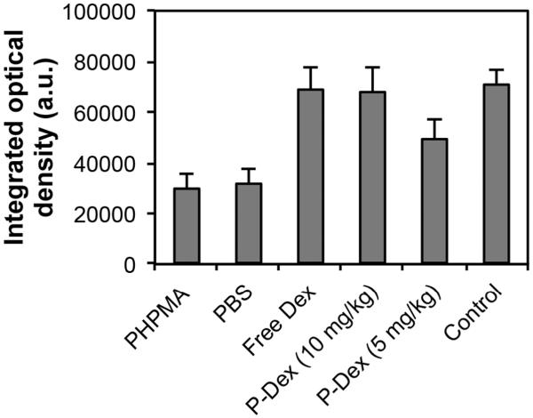
Figure 7.
Quantitative analysis of the peri-implant bone collagen after different treatments. A modified trichrome staining procedure was used to stain the sections. The presence of bone collagen (indicated by the blue color) was quantified for integrated optical density (IOD) using Image Pro Plus software. No significant difference was found when comparing free Dex and P-Dex (10 mg/kg) with the control group, suggesting that free Dex and P-Dex (10 mg/kg) can prevent bone collagen loss. PHPMA and PBS did not show any prevention effect, as evidenced by the significant (P < 0.05) differences between PHPMA vs. control and PBS vs. control.

