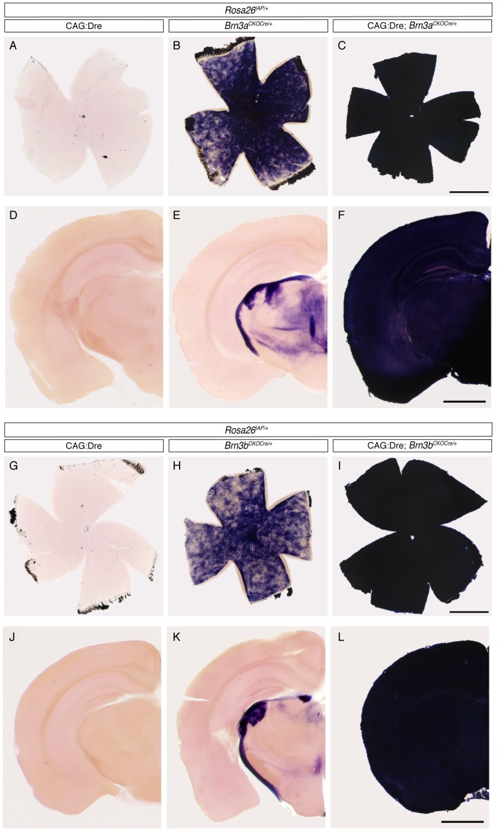
Figure 3. Sequential Dre to Cre recombination suggests ubiquitous Cre expression from the Brn3a and Brn3b loci.
A–C, G–I, Adult retina flat mounts and D–F, J–L, hemispheres from coronal brain sections of mice with indicated genotypes. Note that, in CAG:Dre; ROSA26iAP mice, germline Dre expression does not result in induction of AP positivity from the Cre dependent ROSA26iAP locus (A, D - n = 4; G, J – n = 6). However, Brn3CKOCre; ROSA26iAP tissues show a reduced level of mosaic recombination in RGCs, and corresponding projection areas in the brain (B, E - n = 9; H, K – n = 13). In contrast, tissues from CAG:Dre; Brn3CKOCre; ROSA26iAP mice (C, F - n = 4; I, L – n = 13) show complete conversion to AP positivity, suggesting that the sequential Dre to Cre to AP recombination happened in the totality (or a vast majority) of the tissue. Scale bars in C, F, I and L are 1 mm.

