
Fig. 1.
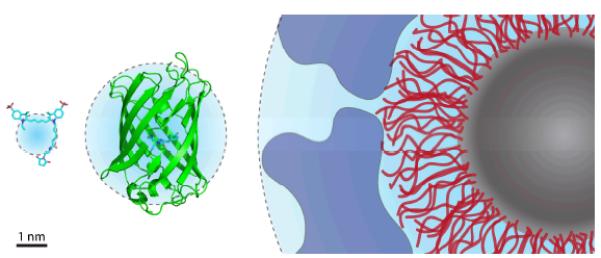
Size comparison of extrinsic fluorophores. From left to right: the organic fluorophore Cy5 (maleimide conjugate), green fluorescent protein, and a quantum dot coated with a passivating polymer layer (red) and a bioconjugating molecule layer (blue). Cyan spheres represent hydrodynamic radii.18, 38, 39