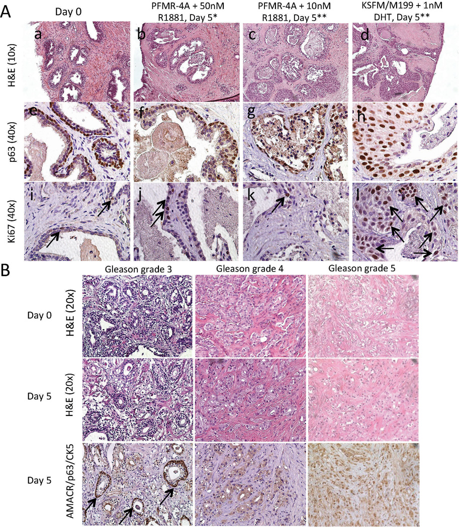
Figure 2.
Histology of benign and PCa TSCs. (A) H&E (a – d) and IHC for p63 (e – h) and Ki67 (i – l, arrows indicate positive nuclei) showed benign TSCs undergoing luminal cell degeneration and/or basal cell hyperproliferation compared to the native tissue (“Day 0”) when cultured in PFMR-4A with 10 nM R1881 (c, g, k) or KSFM/M199 with 1 nM DHT (d, h, l), but not in PFMR-4A with 50 nM R1881 (b, f, j) for 5 days. * = Medium changed every 24 hours. ** = Medium changed every 48 hours. (B) Gleason grades 3, 4, and 5 PCa TSCs cultured in PFMR-4A medium with 50 nM R1881 for 5 days exhibited histologic fidelity to the native “Day 0” tissue as evidenced by H&E staining. AMACR/p63/CK5 staining revealed regions of cancer and some interspersed benign glands (arrows). Representative images were from patient specimens #12 (A), 18, 27, and 30 (B, Table S1).

