Figure 3.
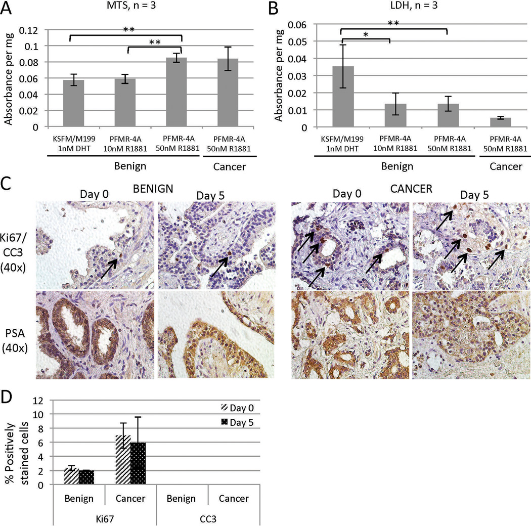
TSC viability and cellular activity. Benign TSCs cultured in PFMR-4A with 50 nM R1881 for 5 days exhibited increased viability by MTS assay (A) and reduced cytotoxicity by LDH assay (B) compared to those cultured under different conditions. PCa TSCs exhibited levels of viability (A) and cytotoxicity (B) similar to those of benign TSCs. ** = p < 0.001, * = p < 0.01. (C) Benign and PCa TSCs cultured for 5 days in PFMR-4A with 50 nM R1881 maintained the same proliferative and apoptotic profiles as the native tissue (“Day 0”) as evidenced by the presence of Ki67-positive cells (arrows) and the absence of CC3 staining, respectively (top row, quantified in D). They also continued to express PSA (bottom row). (D) The percentages of CC3- and Ki67-positive nuclei were quantified from three random 40× fields from each of three tissue slices per condition (average of 97 nuclei per field). Experiments were performed on patient specimens #19, 20, 21 (A, B), 14, and 18 (C).

