
Figure 4.
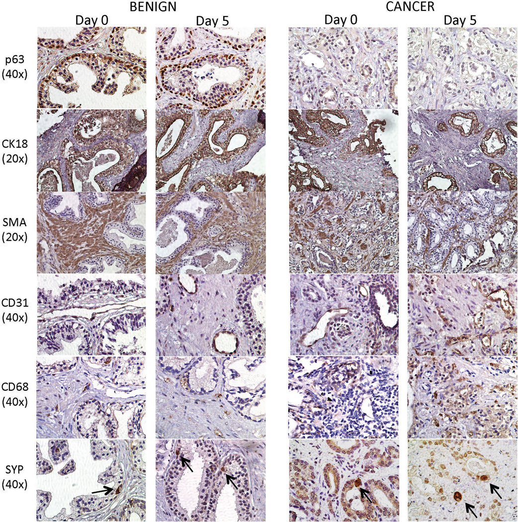
Cellular compositions of benign and PCa TSCs. Cellular markers of basal epithelial cells (p63), luminal epithelial cells and cancer cells (CK18), stromal cells (SMA), endothelial cells (CD31), inflammatory cells (CD68), and neuroendocrine cells (SYP, arrows) were appropriately expressed in benign and PCa TSCs after 5 days in culture. The expression patterns recapitulated those in the native “Day 0” tissues. Representative images were from patient specimens #16, 18, and 27.