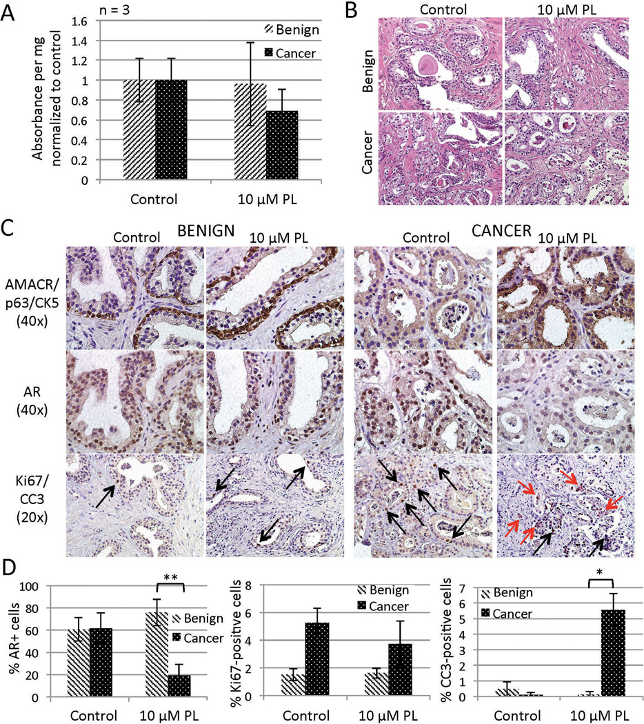
Figure 7.
Responses of TSCs to piperlongumine (PL). PCa but not benign TSCs treated with PL for 6 hr exhibited a non-significant reduction of viability (MTS assay, A) that was mirrored by sporadic regions of luminal degeneration (H&E, B). Further IHC evaluation of benign and PCa TSCs revealed a cancer-specific decrease of AR expression (middle row C, D) and increase of apoptotic cells (red arrows, bottom row C, D). PL treatment did not significantly affect proliferation among benign or PCa cells (black arrows, bottom row C, D). Total nuclei (average of 114 per field) and positively-stained nuclei were blindly counted from three random 40× fields from each of three tissue slices per treatment (D). ** = p < 0.001; * = p < 0.01. Experiments and representative images were from patient specimen #26.

