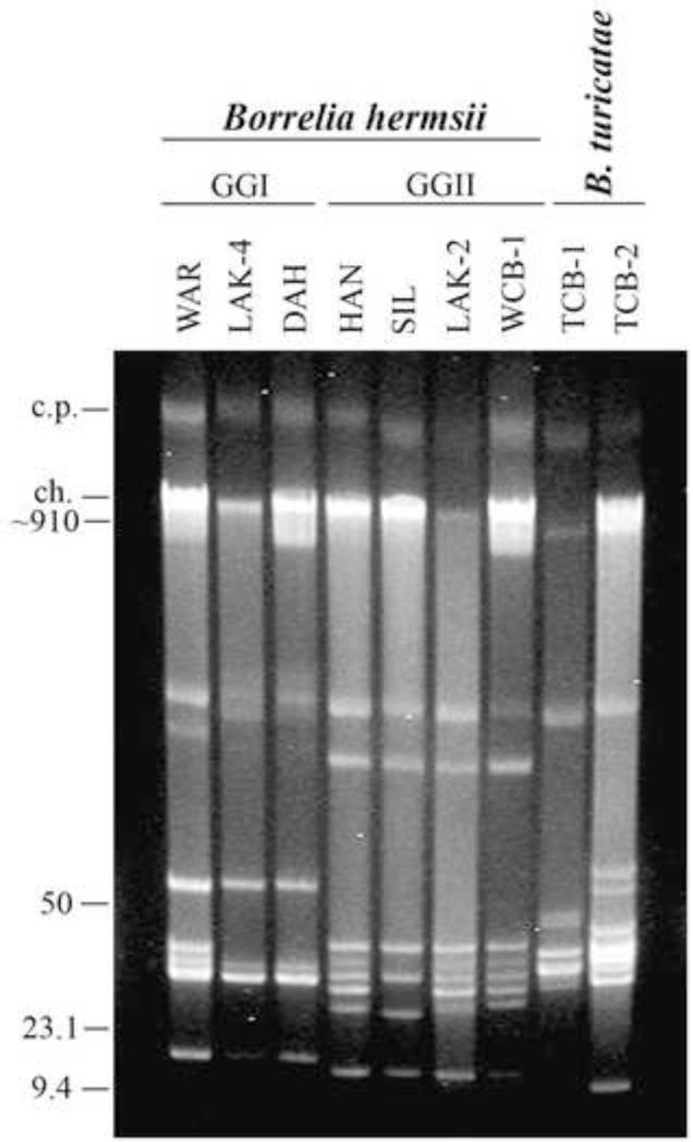
Fig. 3.
Reverse pulse-field agarose gel of uncut genomic DNA from B. hermsii WCB-1 compared to other GGI and GGII isolates and B. turicatae isolated from domestic dogs in Texas. The circular plasmids (c.p.) and linear chromosome (ch.) are shown on the left with estimated DNA sizes shown in kilobases. The remaining bands are linear plasmids of varying sizes.

