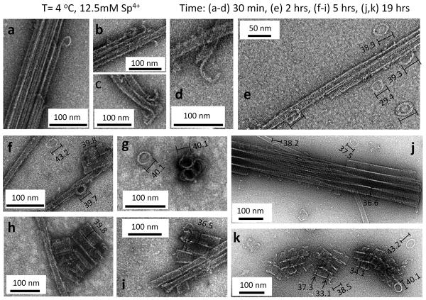
Figure 3. Time-dependent TEM of the pathway of inversion of taxol-stabilized microtubule bundles (BMT) into bundles of inverted tubulin tubules (BITT) at 4 °C and 12.5 mM spermine.
a–d, Transmission electron microscopy (TEM) of early stages of microtubule (MT) bundle disassembly at 30 minutes showing the inside-out curling of protofilaments (PFs) into “pre-ring” structures with a large variation in their diameters (see Supplementary Fig S1). This stage corresponds to Fig. 1c. e, Early-to-intermediate stage TEMs at 2 hrs show the presence of fully formed rings surrounding MT bundles, which dominate the phase at this early-to-intermediate stage where the inverted tubulin structure has not yet formed. TEMs at 1 hr show similar structures. f–i, Intermediate stage TEMs at 5 hrs showing short inverted tubulin tubules (ITTs) and ITT bundles (including an end view of an ITT trimer in g) co-existing with rings, MTs, and MT bundles. j, Late stage TEM at 19 hrs show fully formed bundles of ITTs and few isolated MTs and rings during this late stage. (In the final stage, in the BITT phase, no remaining MTs (and extremely few rings) are found as seen in Fig. 2 both at room T (Fig. 2 b, c) and at 4 °C 24 hrs post addition of 12.5 mM spermine (Fig. 2d).) k, A TEM of a different region of the same sample as in (j) at 19 hrs showing a rare region where short ITTs appear to be forming from the assembly of rings. The variation in the diameter of assembled rings is visible during ITT formation as discussed in the text. The rings in (e–k) have diameters ≈ the diameter of the ITTs. In the measurement of ring size the longer axis was taken because tilts in the ring make it appear as elliptical with the longer axis being a closer estimate of the true diameter. The sample preparations for TEMs (b, c at 30 minutes) and (e–k at 2, 5, 19 hrs) employed a sucrose cushion to remove unpolymerized tubulin after taxol-stabilization of MTs. The sucrose cushion was not employed for TEM samples (a, d at 30 minutes) (see Sample Preparation in the Methods section). All TEMs were at taxol/tubulin molar ratio = 0.55.

