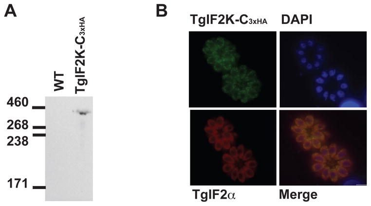
Fig. 2.
TgIF2K-C is expressed in the cytosol ofToxoplasma gondii tachyzoites. (A) TgIF2K-C was endogenously tagged with three tandem hemagglutinin (HA) tags (TgIF2K-C3xHA) and expression was analyzed by western blot of parasite lysate using anti-HA antibody. Protein lysate prepared from the parental line (wild-type; WT) was loaded as a control. Sizes are shown in kilodaltons. (B) IFA using HA-antibody reveals a cytoplasmic distribution for TgIF2K-C3xHA (green). DAPI (blue) was used as a co-stain to highlight the parasite nucleus and total TgIF2α highlights the parasite cytosol (red). Scale bar = 5 um.

