Abstract
Neurofibrillary tangles (NFTs), a hallmark of Alzheimer’s disease (AD), are intracellular silver and thioflavin S-staining aggregates that emerge from earlier accumulation of phospho-tau in the soma. Whether soluble misfolded but nonfibrillar tau disrupts neuronal function is unclear. Here we investigate if soluble pathological tau, specifically directed to the entorhinal cortex (EC), can cause behavioral or synaptic deficits. We studied rTgTauEC transgenic mice, in which P301L mutant human tau overexpressed primarily in the EC leads to the development of tau pathology, but only rare NFT at 16 months of age. We show that the early tau lesions are associated with nearly normal performance in contextual fear conditioning (CFC), a hippocampal related behavior task, but more robust changes in neuronal system activation as marked by Arc induction and clear electrophysiological defects in perforant pathway synaptic plasticity. Electrophysiological changes were likely due to a presynaptic deficit and changes in probability of neurotransmitter release. The data presented here support the hypothesis that misfolded and hyperphosphorylated tau can impair neuronal function within the entorhinal-hippocampal network, even prior to frank NFT formation and overt neurodegeneration.
Keywords: tau, Arc induction, synaptic dysfunction, Alzheimer’s disease
Introduction
Neurofibrillary tangles (NFTs), intracellular aggregates of misfolded and hyperphosphorylated tau protein, are a neuropathological feature of Alzheimer’s disease (AD) and other tauopathies. In AD, NFTs correlate well with amounts of phospho-tau immunoreactivity, synapse loss, and neuronal loss. Each of these markers correlate well with dementia severity [21] so that the contribution of NFTs or phospho-tau, as opposed to, for example, neuronal or synaptic loss, cannot be discerned. Distinguishing the roles of soluble and fibrillar tau is equally difficult. Indeed, recent studies suggest that more soluble forms of tau, rather than fibrillar tangles, may be involved in neuronal dysfunction [28,63,44,50,45,29]. We now take advantage of a recently developed mouse model with focal tau expression largely limited to the entorhinal cortex (EC) to examine the consequences of the accumulation of soluble tau and evaluate its effects on neural system integrity at a time point prior to anatomical neurodegenerative changes.
The layer II entorhinal cortex (EC-II) neurons that project to the hippocampus, via the perforant pathway (PP), are critical for memory function. They also are the first cortical neurons to be affected by NFTs in AD [4,20]. In the present study we examine the recently characterized rTgTauEC transgenic mouse in which P301L human mutant tau is overexpressed primarily in the EC, leading to pathological tau inclusions in the EC-II as animals age [9]. These animals thus mimic, from an anatomical perspective, the types of lesions that occur in very early AD and provide a platform to test the hypothesis that development of pathological tau in the EC, at a pre-tangle stage, results in memory deficits or synaptic dysfunction. We found that restricted accumulation of pathological tau in the EC and perforant pathway, prior to tangles, synaptic loss, or neuronal loss, allows nearly normal performance on hippocampal related behavior tasks, but more marked changes in hippocampal neural system activation as indicated by Arc induction and defects in hippocampal electrophysiological properties. These observations implicate disturbance of synaptic transmission and plasticity in the perforant pathway at an age prior to the development of fibrillar tau aggregates (i.e. NFTs), synaptic or neuronal loss, providing evidence that favors the idea that soluble, misfolded tau can impact neural system function.
Materials and Methods
Animals
rTgTauEC mice: We generated transgenic animals (called rTgTauEC – for reversible tau restricted to entorhinal cortex) by crossing FVB-Tg(tetO-TauP301L) 4510 mice [47] with a transgenic mouse line on a C57BL/6 genetic background expressing tetracycline transactivator under the control of the Klk8 neuropsin promoter (EC-tTa) that was developed at the Scripps Research Institute [62]. F1 offspring were used as experimental animals ensuring a uniform 50:50 mix of FVB and C57BL/6 genetic background. Inheritance of both the responder and activator transgenes (designated rTgTauEC) results in P301L mutant tau expression constrained to layer II of the EC and pre and para subiculum. Notably, the restricted expression of the transgene has been characterized by three independent groups [9,35,18]. The limited anatomical expression of the transgene was unquestionably confirmed by using definitive laser capture microdissection and RT-PCR that the tau mRNA is not detectable in the DG of rTgTauEC mice [9].
Age matched littermates expressing only the activator transgene were used as human taunegative controls. rTgTauEC and control mice were identified by PCR screening using the primer pairs 5’-ACCTGGACATGCTGTGATAA-3’ and 5’-TGCTCCCATTCATCAGTTCC-3’ for activator transgenes [62], and 5’-TGAACCAGGATGGCTGAG CC-3’ and 5’-TTGTCATCGCTTCCAGTCCCCG-3’ for responder transgenes [47,9]. All animal experiments were performed in accordance with national guidelines (National Institutes of Health) and approved by Massachusetts General Hospital and McLaughlin Institute Institutional Animal Care and Use Committees.
Immunohistochemistry
Standard immunofluorescence techniques were used. Briefly, animals were sacrificed by CO2 inhalation and brains were flash-frozen in M-1 mounting medium (Shandon, Thermo Scientific) horizontal cryostat sections were cut at 10 m, sections were fixed in 4% paraformaldehyde for 10 min, permeabilized by 20 min incubation in 0.1% Triton solution. After blocking in 5% normal goat serum (NGS) for 1 hour, the appropriate primary antibody was applied in 5% NGS, and sections were incubated overnight at 4°C. The antibody Tau13 which recognizes the amino acid residues 20–35 on the longest isoform of human tau [2,25] (Covance; 1:1,000) was used to detect human tau; the conformation-specific Alz50 and MC1 antibodies (courtesy Peter Davies, Albert Einstein College of Medicine; 1:50) [30,5,3] used to detect misfolded tau, and PHF1 (pSer396/404) [17,39] (courtesy Peter Davies, Albert Einstein College of Medicine; 1:500) and MC1 [23,57] (courtesy Peter Davies, Albert Einstein College of Medicine; 1:200) or Gallyas silver staining was used to detect neurofibrillary tangles. Sections were washed and incubated with Fluorescent Alexa Fluor 488 (Jackson ImmunoResearch; 1:250) secondary antibody in 5% NGS for 1 hr at room temperature and stained with DAPI. (1:1,000).
Gallyas Silver Staining
Gallyas silver staining was performed as described previously [15]. Images for figures were collected on an upright Olympus BX51 microscope (Olympus America, Center Valley, PA) with a 40× objective.
Densities of human tau, Alz50 and Gallyas stained cells
The layer II of the EC and the DG were outlined on five sections per mouse (n=3 per group) using Image J. Tau13, Alz50 and Gallyas positive neurons were counted and the density of EC or DG neurons positive for each marker was calculated by dividing the number of total counted neurons by the size of the area outlined (EC or DG) in each section. The five sections from each animal were averaged for the mean value per animal. Finally, the mean value of all animals was calculated and values are presented as mean ± SEM.
Western Blot Analysis
Western blot analysis was performed as described previously [9] Brains were homogenized in RIPA buffer (Invitrogen) supplemented with a cocktail of protease and phosphatase inhibitors (Roche). Samples were homogenized using a Polytron and the protein content was determined by BCA protein assay (Thermo Scientific). The materials for SDS-PAGE were obtained from Invitrogen (NuPAGE system). Protein lysates were boiled in sample buffer consisting of lithium dodecyl sulfate sample buffer and reducing agent and resolved on 4%–12% Bis-Tris polyacrylamide precast gels in MES SDS running buffer. 30 g of protein were loaded per lane, proteins were transferred onto nitrocellulose membranes Protran (Whatman) in transfer buffer containing 20% methanol. Blots were blocked in Odyssey blocking buffer (Li-Cor biosciences), followed by incubation with primary antibodies (-actin [mouse monoclonal antibody, Sigma; 1:10,000]; K9JA phosphorylation-independent pan-tau antibody [rabbit polyclonal antibody, Dako; 1:20,000] [56], and detected with anti-mouse or anti-rabbit IgG conjugated to IRDye 680 or 800 (Li-Cor Biosciences; 1:10,000). Western blots were developed using the Odyssey ® Imaging System (LI-COR Biosciences), which allows detection of multiple primary antibodies probed on the same blot. Densitometric and MW analyses were performed using ImageJ software (National Institutes of Health). Band density values were normalized to - actin levels. Mean band densities for samples from rTgTauEC mice were normalized to corresponding samples from 24 month-old mice that did not receive doxycycline treatment.
Sarkosyl Insolubility Assay
Extraction of sarkosyl-insoluble tau was performed as previously described [19]. Briefly, whole frozen brains of 24 and 16-month-old rTgTauEC mice were homogenized by polytron in 10 volumes of buffer H (10 mM Tris-HCl [pH 7.5] containing 0.8M NaCl, 1 mM EGTA, and 1 mM dithiothreitol) and spun at 100,000 × g for 30 min at 4°C. Another 2 ml of buffer H was added to the pellet and the samples were homogenized again by polytron, incubated in 1% Triton X-100 at 37°C for 30 min. Following the incubation, the samples were spun at 100,000 × g for 30 min at 4°C, the pellet was homogenized by polytron in 1 ml of buffer H and was then incubated in 1% sarkosyl at 37°C for 30 min and spun at 100,000 × g for 30 min at 4°C. The supernatant was then collected (sarkosyl-soluble fraction). Detergent-insoluble pellets were extracted in 100 µl of urea buffer (8 M urea, 50 mM Tris-HCl [pH 7.5]), sonicated, and spun at 100,000 × g for 30 min at 4°C. The supernatant was then collected (sarkosyl-insoluble fraction).
Behavior Experiments
Behavioral tests were performed at the NeuroBehavior Laboratory Core at Harvard NeuroDiscovery Center. The experimenter was blind to genotype for all behavioral tests. For detailed methods see Supplemental Methods.
Electrophysiology
Experiments were performed as previously reported with modifications [41,6,31]. Animals were deeply anesthetized using isoflurane and killed by decapitation. The brain was quickly removed and the brain was dissected in an ice-cold cutting solution. Acute horizontal slices (400 µm) were prepared from non-transgenic controls and rTgTauEC mice at 16 months of age. Slices were cut on a DTK-2000 microslicer (Dosaka) in ice-cold extracellular cutting solution containing the following (in mM): 215 sucrose, 2.5 KCl, 20 glucose, 26 NaHCO3, 1.6 NaH2PO4, 1 CaCl2, 4 MgCl2, and 4 MgSO4. Slices were then placed, at room temperature, in a holding chamber containing 50% cutting solution and 50% artificial cerebral spinal fluid (CSF) (ACSF) recording solution containing the following (in mm): 124 NaCl, 26 NaHCO3, 10 glucose, 2.5 KCl, 1 NaH2PO4, 2.5 CaCl2, and 1.3 MgSO4. After 30 min, the 1:1 solution was switched to ACSF. The slices were kept at room temperature for at least 1 hr before being transferred to the recording chamber. Cutting and recording solutions were both saturated with 95% O2 and 5% CO2, pH 7.4. Experiments were performed at room temperature. A patch-type pipette filled with 1 M NaCl was used to record field excitatory postsynaptic potentials (fEPSPs) and were evoked every 20 sec with a bipolar tungsten electrode in the presence of 100 µM picrotoxin (Sigma-Aldrich) to block inhibitory transmission.
Data was acquired with Digidata-1322A and digitized at 10 kHz using the pClamp 10 software. Recordings were performed with a MultiClamp 700A amplifier (Axon Instruments), and output signals were filtered at 3 kHz. The data was analyzed using MiniAnalisys (Synaptosoft). The stimulating and recording pipettes were positioned at the same depth in the slice and the distance between them was kept constant (~200 µm). Input/output (I/O) curves were generated using stimulus intensities from 10 to 100 µA in increments of 10 µA and plotted as a function of fiber volley amplitude. Paired-pulse facilitation and depression were assessed by using 50 and 100 ms inter-stimulus intervals. Before LTP induction, stable baseline responses were acquired for at least 20 min. LTP was induced at baseline stimulus intensity using high-frequency stimulation (HFS) which consisted of four trains of 50 pulses at 100 Hz with 10 sec inter-train intervals. The magnitude of LTP was estimated by comparing averaged responses 15–45 min after induction with baseline-averaged responses before induction. For statistical analysis, responses were collected and every 3 responses (1 min periods) for the LTP experiments or every five responses for the I/O and PPD/PPF experiments were averaged. fEPSP slope or amplitude were calculated. As previously described, fEPSP were evoked and recorded at CA3-CA1 and PP-DG [18]. For more detailed electrode placing information see Supplemental material.
Real Time Quantitative RT-PCR analysis of Arc mRNA
Arc mRNA levels were measured 30 min after rTgTauEC and non-transgenic control mice were re-exposed to CFC (or not exposed) (n=5 or 6 per group). RT-PCR procedure was performed as previously reported with modifications [52]. Animals were sacrificed, the brains were removed and the hippocampus was dissected. Total RNA was extracted from hippocampus by homogenizing tissue in 500 l of Trizol reagent (Invitrogen) followed by addition of 100 l chloroform at room temperature and incubated for 10 min. Samples were centrifuged for 15 min at 12,000g at 4°C and the aqueous phase was transferred to new, RNase-free Eppendorf tubes. The RNA was precipitated by addition of 250 l of isopropanol and frozen for 1hr at −80°C. These samples were then centrifuged for 15 minutes at 12,000g at 4°C, the RNA pellets were washed in 70% EtOH, air-dried for several minutes, and then suspended in 14 l of UltraPure™ DNase/RNase-Free Distilled Water. RNA samples were assayed for quality with an Agilent 6000 Bioanalyzer and a Nanodrop spectrophotometer. Reverse transcription was carried out on all RNA samples (Superscript II, Invitrogen) and random hexamers. The relative amount of Arc was normalized to the level of internal control housekeeping gene message for glyceraldehyde-3-phosphate dehydrogenase (GAPDH) mRNA levels. Real-time PCR was performed according to the comparative threshold cycle (CT) method (SmartCycler manufacturer's instructions). Differences in threshold crossing cycle between Arc and GAPDH (equal to DeltaCT) were calculated for each condition; then the levels of Arc expression were computed as Delta-Delta-CT, and Arc mRNA levels in CFC exposed and unexposed transgenic and non-transgenic animals were calculated and normalized to non-transgenic unexposed mice. qPCR analysis (on Bio-Rad iCycler) of the cDNA product was carried out using primers against Arc forward, 5'-GAAGGAGTTTCTGCAATACAGTGAG-3'; and reverse, 5'-ACATACTGAATGATCTCCTCCTCCT-3' and GAPDH forward, 5'-ATGACATCAAGAAGGTGGTG-3' and reverse, 5'-CATACCAGGAAATGAGCTTG-3'. Duplicates or triplicates of cDNA samples were added to a 25 l reaction containing 12.5 l SYBR green Mastermix (Applied Biotechnology).
Statistical analyses
Normality of data was assessed with a Shapiro–Wilks test. Data were analyzed with appropriate statistics including Student's t test, Mann-Whitney test, one-way ANOVA followed by Tukey post hoc test (when necessary) and Kolmogorov-Smirnov test (KS-test). P values <0.05 were considered to be statistically significant. Data were reported as mean ± SEM.
Results
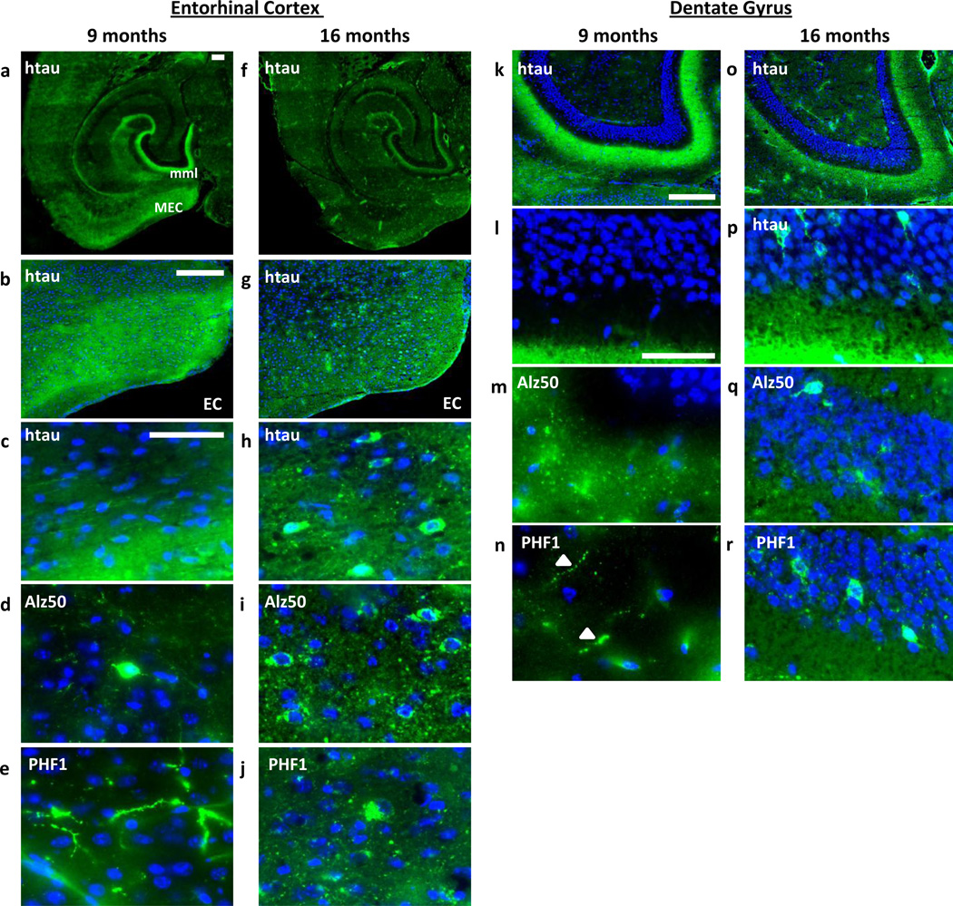
To assess tau pathology in rTgTauEC mice we immunostained horizontal brain slices from 9 and 16 month-old animals with different tau antibodies. At 9 months of age, human tau protein (htau) assessed by the human tau specific antibody Tau13 (that shows no reactivity in control mouse brain, Fig S1a), has a restricted distribution of htau in the medial entorhinal cortex (MEC) and in the axon terminals in the middle molecular layer (mml) of the dentate gyrus (DG), which receives axons originating in the MEC (Fig 1a–e; Table 1). At this age only a very small number of Alz50-positive (misfolded tau and a pre-tangle marker) cell bodies (Fig 1d; Table 1) and Alz50 and PHF1-positive (hyperphosphorylated tau) neurites (Fig 1d and 1e) were observed in the EC. The Alz50 staining pattern was confirmed using a different misfolded tau antibody MC1 (FigS1b–c). By contrast, 16 month-old rTgTauEC mice show numerous aggregates positive for human tau, Alz50 and PHF1 in the EC cell bodies (Fig 1f–j). The phenotype of “spreading” of tau to the DG granule cells occurs largely in older mice (21–24 months), although even at 16 months rare granule cells were positive for human tau, Alz50 and PHF1 (Fig 1o–r). Axons and dendrites, but no soma, were human tau immunopositive in the hippocampus at 9 months of age (Fig 1k–n). Thus 16 month-old animals show a different pattern of tau distribution (Fig 1a–b vs f–g), and the levels of tau are considerably increased (Fig S1d), consistent with previous reports [9].
Fig. 1.
Distribution of tau in the entorhinal cortex (EC) and dentate gyrus (DG) of rTgTauEC mice at 9 and 16 months of age. EC: a and f Low magnification view of medial horizontal sections from 9 and 16 month-old rTgTauEC mouse brains showing the distribution of human tau using the human tau specific phosphorylation-independent Tau13 antibody. At 9 months, human tau (htau) has a restricted distribution in the MEC and in the axonal terminals in the middle molecular layer (mml) of the DG a (b and c higher magnifications of the EC). 16 month-old rTgTauEC mice show numerous human tau positive cell bodies in the EC f (g and h higher magnifications of the EC). Alz50 staining shows rare Alz50-positive EC cell bodies at 9 months d and a number of Alz50-positive neurons in the EC at 16 months i. Immunohistochemistry using PHF1 shows PHF1-positive neurites in the EC at 9 months e and PHF1-positive aggregates in the EC neurons of 16 month-old rTgTauEC mice j. DG: At 9 months, human tau k–l, Alz50 m and PHF1 n staining are limited to mml of the DG. At 16 months, a few granule cells are positive for human tau o–p, Alz50 q and PHF1 r. Tau is shown in green and DAPI nuclei are shown in blue. Scale bars 200 m (a–b, f–g, k–r) and 50 m (c–e, h–j). White arrows indicate axonal tau staining.
Table 1.
Densities of human tau, Alz50 and Gallyas stained cells in entorhinal cortex and dentate gyrus of 16 month-old rTgTauEC mice (n = 3 mice). Results are expressed as the mean ± SEM.
| Entorhinal Cortex | |
|---|---|
| number neurons /mm2 | |
| human tau | 85.22 ± 7.70 |
| Alz50 | 78.68 ± 8.91 |
| Gallyas | 9.1 ± 2.20 |
| Dentate Gyrus | |
| number neurons /mm2 | |
| human tau | 58.96 ± 22.08 |
| Alz50 | 46.93 ± 2.49 |
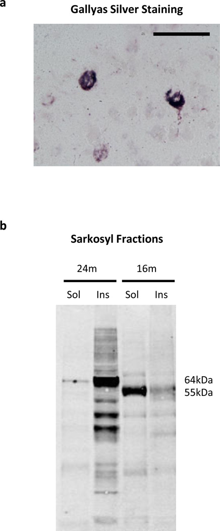
The lesions present in the EC at 16 months of age were primarily still pre-tangle in their staining properties. There were only rare Gallyas silver positive neurons in the EC with the density of 9.1 neurons/mm2 (on the order of 0–5 neurons per slice) (Fig 2a; Table 1) and no thioflavin S-staining suggesting that the vast majority of the prominent tau positive neurons are in a pre-tangle state. As the animals age, the disease progresses and at 24 months of age the density of Gallyas positive tangles in the EC increases dramatically to 1,775± 534 Gallyas-positive neurons/hemisphere or 26 ± 8.76 number neurons /mm2. Gallyas positive neurons in the dentate gyrus at 24 months of age were infrequent, usually <5 per slide. Consistent with pre-tangle pathology, immunoblotting of sarkosyl fractions from 16 month-old mice rTgTauEC mice revealed very low levels of sarkosyl insoluble tau species at 55 kDa with most tau detected in the soluble fraction as a 55 kDa species. A 64 kDa insoluble hyperphosphorylated tau species that could be detected at 24 months [9] was absent in 16 month-old animals brains (Fig 2b).
Fig. 2.
Rare Gallyas silver positive tangles in the EC a. rTgTauEC animals rarely show sporadic silver positive neurofibrillary tangles in the EC at 16 months of age. Scale bar 50 µm. b. Immunoblotting of sarkosyl fractions from rTgTauEC mice using a K9JA total tau antibody revealed very low levels of sarkosyl insoluble tau species at 55 kDa in 16 month-old mouse with most of tau detected in the soluble fraction as a 55 kDa species of tau. A 64 kDa insoluble hyperphosphorylated tau species detected at 24 months but absent in 16 month-old animals brains.
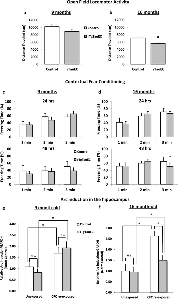
The EC-hippocampus system plays a critical role in memory formation and memory consolidation. We tested the hypothesis that accumulation of tau in the perforant pathway might be associated with cognitive decline. We used the open field test to measure the general locomotor and exploratory activity of rTgTauEC mice. While 9 month-old transgenic mice showed similar total distance traveled compared to age-matched non-transgenic controls (Fig 3a), at 16 months transgenic mice showed a significant decrease (~20%) in total distance traveled (p=0.014) (Fig 3b). There were no changes in the percent distance traveled in the center (Fig S2a) or in the number of vertical counts (Fig S2b) suggesting no changes in anxiety levels or exploratory behavior. The 16 month-old animals showed similar latency to fall on the rotarod test compared to controls, consistent with normal motor function (Fig S2c). Moreover, 16 month-old animals performed similarly to controls on the radial arm maze, a test dependent on the hippocampal formation, showing intact spatial memory (Fig S2d).
Fig. 3.
a. Open field locomotor and exploratory activity test in rTgTauEC mice revealed no changes in the activity of 9 month-old transgenic mice (control, n = 12; rTgTauEC, n = 12) and a significant decrease (~20%) in the total distance travel (p=0.014) of transgenic mice at 16 months of age (control, n = 6; rTgTauEC, n = 14) compared to age-matched non-transgenic controls b. Contextual fear conditioning (CFC) shows no changes in fear response measured by freezing in 9 month old mice (24 hrs: control, n = 6; rTgTauEC, n = 14; 48 hrs: control, n = 3; rTgTauEC, n = 6) c but 16 month-old rTgTauEC mice (24 hrs: control, n = 12; rTgTauEC, n = 12; 48 hrs: control, n = 4; rTgTauEC, n = 6) had a small decreased freezing time (48 hrs) compared to age-matched controls (p=0.0414) d. Activation of Arc mRNA expression induced by hippocampus-dependent fear response to contextual fear conditioning (CFC) was assessed using qRT-PCR. Mice that were re-exposed to CFC 48 hrs after training and sacrificed 30 minutes after re-exposure were compared to unexposed mice. e 9 month-old controls and transgenic mice re-exposed to CFC show a significant increase in Arc mRNA levels in the hippocampus compared to unexposed mice. There was no difference in Arc mRNA levels of rTgTauEC mice compared to controls (9 months: control, unexposed n = 4, re-exposed n=5; rTgTauEC, unexposed n = 6, re-exposed n = 6). f 16 month-old transgenic mice have significantly less Arc activation following CFC re-exposure compared to controls. (16 months: control, unexposed n = 3, re-exposed n=4; rTgTauEC, unexposed n = 3, re-exposed n = 6). Results are expressed as means + SEM. Comparisons between groups were made by t test or Mann-Whitney test (for CFC). * indicates statistical significance, p < 0.05. For CFC: * indicates statistically significant difference between unexposed and re-exposed mice, p < 0.05. # indicates statistically significant difference between control and transgenic mice, p < 0.05. n.s. indicates no statistical significance.
Contextual fear conditioning (CFC) also depends on the limbic system and represents a form of associative learning in which the animal learns to predict aversive events. We assessed the fear response of rTgTauEC mice using a CFC paradigm consisting of three days of testing with training (shock delivery) performed on the first day, followed by two additional days of testing (24 hrs and 48 hrs after training) (Fig S2e). We found that there were no changes in fear response measured by freezing in 9 month rTgTauEC mice compared to controls (Fig 3c). While 16 month-old rTgTauEC had a mild decreased freezing time during the last minute on the second day of testing (48 hrs) (Fig 3d), this represents a mildly effect with a small sample size (control, n = 4; rTgTauEC, n = 6), suggesting nearly normal CFC behavior. There were no differences in the freezing time at baseline (prior to shock delivery) between controls and transgenic mice in either age group (Fig S2f and S2g).
Next we assessed whether tau accumulation leads to changes in the activation of the plasticity-related activity regulated, cytoskeletal-associated gene Arc, which is induced in hippocampus-dependent fear conditioning [8,40]. Both controls and transgenic mice that had been re-exposed to CFC were compared to unexposed mice. We quantified Arc mRNA by quantitative real-time polymerase chain reaction (qRT-PCR) in the hippocampus 30 minutes after CFC behavioral experience. Half the animals that were tested on the CFC task at 24 hrs after training were re-tested at 48 hrs and sacrificed 30 minutes thereafter. mRNA levels were determined in CFC re-exposed and CFC unexposed mice. As expected, in 9 month-old mice there was a significant increase in Arc mRNA levels in the hippocampus of both controls (p=0.019) and rTgTauEC transgenic (p=0.0087) mice that had been re-exposed to CFC compared to unexposed mice (Fig 3e) showing the expected Arc induction in response to CFC. No difference in the pattern of Arc mRNA levels of rTgTauEC mice compared to controls was observed in animals at 9 months of age, consistent with the observed behavioral results (Fig 3e). By contrast, 16 month-old transgenic mice showed significantly less Arc activation following CFC re-exposure compared to controls (Fig 3f) (p=0.0254). At 16 months, both controls and rTgTauEC transgenic mice that had been re-exposed to CFC showed a significant increase in Arc mRNA levels in the hippocampus compared to unexposed mice (p=0.0286 and p=0.0223, respectively), but the size of the increase of Arc transcription was significantly smaller in the transgenic animals by ~57%.
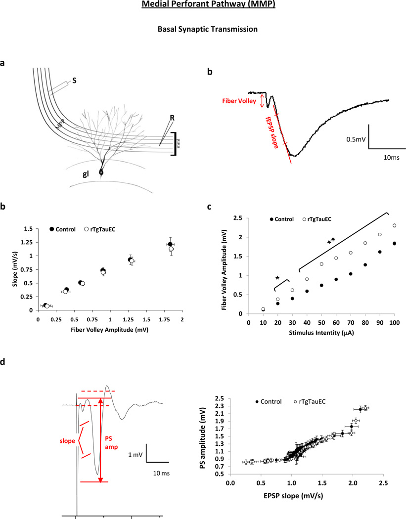
These results suggest subtle that changes in one of the two hippocampal dependent memory tests assessed in this study as well as changes in the responsiveness of the neural system activity marker Arc occur in the rTgTauEC model as they age. To further explore whether human mutant tau overexpression in the EC and perforant pathway impairs synaptic function in the hippocampus, we measured baseline synaptic transmission, short-term plasticity and longterm potentiation (LTP) at the EC medial perforant pathway to dentate gyrus granule cell synapses (PP-DG) in 16 month-old mice as illustrated by the schematic diagram of the medial perforant pathway with the positioning of the stimulating (S) and recording (R) electrodes (Fig 4a). Recordings were also performed at the Schaffer collateral synapse to CA1 synapse (CA3-CA1) which we anticipated would be unaffected by EC specific human mutant tau overexpression. We recorded extracellular field excitatory postsynaptic potentials (fEPSP) from horizontal EC-hippocampal slices. To assess baseline synaptic transmission strength we examined the input-output relationship of PP-DG and CA3-CA1 synapses in response to single electrical stimuli and found no significant difference between transgenic and control animals at either synapse when the fEPSP was plotted against the fiber volley amplitude (Fig 4b and Fig S3b). There was increased presynaptic volley amplitude in the PP-DG synapse of rTgTauEC mice compared to controls when the fiber volley amplitude was plotted as a function of the stimulus intensity (Fig 4c), suggesting enhanced axonal excitability and indicative of a presynaptic alteration.
Fig. 4.
Synaptic transmission at the EC medial perforant pathway to dentate gyrus granule cell synapses (PP-DG). Extracellular field excitatory postsynaptic potentials (fEPSP) were recorded from horizontal EC-hippocampal slices of 16 month-old rTgTauEC and age-matched nontransgenic controls. a Schematic diagram of medial perforant pathway (MPP) synapto-architecture showing middle molecular layer (mml) and granule cells (gl) of the dentate gyrus with the positioning of stimulation (S) and recording (R) electrodes. b Typical shape of a fEPSP illustrating the method of assessment of presynaptic fiber volley amplitude and fEPSP slope. b Basal synaptic transmission was examined by building input-output (I/O) relationship at the PP-DG synapse in response to single electrical stimuli ranging from 10 to 800 A and plotted as the relationship between fEPSP slope and fiber volley amplitude, showing no significant difference between transgenic mice compared to control animals. c Fiber volley amplitude was plotted as a function of stimulus intensity and it shows a significant increase in fiber volley amplitude for a given stimulus intensity in the rTgTauEC mice compared to controls. (n= [control, n = 10 (3); rTgTauEC, n = 8 (4)]). d Typical shape of a population spike illustrating the method of assessment of amplitude of the population spike and the slope of the EPSP. E-S coupling was assessed by measuring the slope of the initial population EPSP and the amplitude of the population spike. Curves were generated for EPSP slope vs. population spike amplitude. There was no change in E-S coupling in rTgTauEC mice compared to controls the field suggesting that these animals do not have a post-synaptic change. (n= [control, n = 4 (3); rTgTauEC, n = 4 (3)]).
To access if there are post-synaptic changes in the PP-DG synapse associated with enhanced pre-synaptic axonal excitability, we measured the slope of the initial population EPSP and the amplitude of the population spike as described [59]. This measurement can be used as an indicator of post-synaptic changes since more excitable cells fire action potentials with lower amplitude synaptic input. There was no consistent shift of the EPSP slope vs. population spike amplitude curve (E-S coupling) suggesting that these animals do not show a post-synaptic change that can be detected by this measurement (Fig 4d).
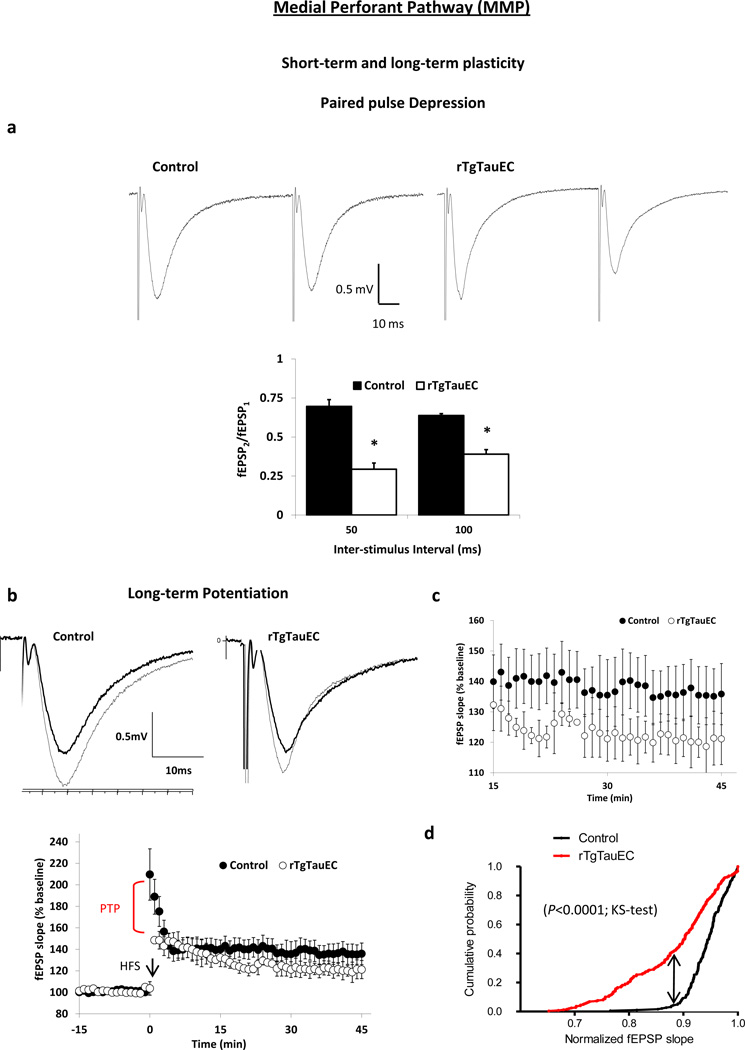
Next we measured paired pulse ratio (PPR) in the PP-DG and CA3-CA1 synapses, a form of short-term plasticity traditionally used as an indirect way to estimate the probability of transmitter release (Pr). PPR is inversely correlated with Pr; synapses with low Pr usually show paired pulse facilitation (PPF), whereas synapses with high Pr show paired-pulse depression (PPD) [36,53]. We measured PPR in the PP-DG synapse that normally shows PPD, such that a single stimulus leading to synaptic activation will depress a subsequent synaptic response (Fig 5a). We used two inter-stimulus intervals (ISI) (50 and 100ms) with equal intensity for both stimuli. We used stimulus intensity that elicits similar initial fEPSP amplitude in slices from controls and transgenic ranging from 0.9 to 1.5mV. In slices from transgenic mice, PPD was significantly increased compared to controls, as measured by the PPR (ratio between the amplitude of the second pulse and the first pulse) (p= 0.0062 for 50ms; p= 0.0007 for 100ms). This suggests an increase in the probability of transmitter release in the transgenic mice, consistent with a presynaptic alteration (Fig 5a).
Fig. 5.
Synaptic plasticity at the medial perforant pathway. a Short-term plasticity was evaluated by measuring paired pulse depression (PPD) of the PP-DG synapse. Paired-pulse ratios (PPRs) calculated as the ratio between the amplitude of the second response (fEPSP2) and the first response (fEPSP1) to two pulses delivered either 50ms or 100ms apart. Representative fEPSP for responses with 50ms interval is shown. There was a significant increase in PPD shown by decreased PPR (n= [control, n = 6 (3); rTgTauEC, n = 6 (4)]). b–d LTP was evaluated by comparing fEPSP slopes as a percentage of the pre-tetanus baseline fEPSP slopes (n= [control, n = 6 (3); rTgTauEC, n = 8 (4)]). Representative traces before (bold line) and 30–45 min after (thin line) the induction. There was a significant reduction in the post-tetanic potentiation (PTP) after high-frequency stimulation (HFS) (d). fEPSP responses between 15 to 45 min after HFS are shown in E. Results are expressed as means ± SEM. * indicates statistical significance, p < 0.05 (b–e). Comparisons between groups were made by t test (b–e). f Cumulative probability distribution plots for recordings from 15 to 45 min after LTP induction show a significant decrease in LTP elicited in rTgTauEC mice compared to controls. Comparison between groups was made by Kolmogorov-Smirnov test (KS-test). * indicates statistical significance, p < 0.05. (n = [number of slices (number of animals)]).
In contrast to the PP-DG synapse that shows PPD, CA3-CA1 synapses exhibit PPF, such that a single synaptic activation facilitates a subsequent synaptic response. We used 50 and 100ms ISI and initial amplitude responses ranging from 1.6 to 2 mV. We measured the PPR at the CA3-CA1 synapse and found no changes in PPF in transgenic mice compared to age-matched controls (Fig S3c), as expected because the transgene is not expressed in this circuit.
In addition, we assessed whether long-term potentiation (LTP) is altered in the medial perforant pathway rTgTauEC mice (Fig 5b–d). LTP is a long-lasting strengthening of the response of postsynaptic neurons to high-frequency stimulation and is widely accepted as a critical molecular mechanisms that underlies learning and long-term memory. Post-tetanic potentiation (PTP) is a form of short-term plasticity generally thought to be presynaptic with a mechanism similar to PPF and PPD, in that after high frequency stimulations, often used for LTP induction, there is a high level of residual calcium in the presynaptic cell, temporarily increasing the probability of release. LTP was induced after the acquisition of a stable baseline. Baseline fEPSP slope and fiber volley amplitude were kept similar for slices from controls and transgenic mice with fEPSP slope ranging from 0.9 to 1.1 mV/ms and fiber volley 0.75 to 0.95 mV. Slices from transgenic animals demonstrated a significant reduction in PTP after high-frequency stimulation in the PP-DG synapse (p<0.0001) (Fig 5d). Responses reached a steady state by 15 minutes after tetanus (Fig 5e). Cumulative probability plots of response amplitudes 15 to 45 minutes following high frequency stimulation of the medial perforant pathway revealed that LTP was markedly decreased in transgenic mice compared to control mice (p<0.0001) (Fig 5f). In contrast, tetanic stimulation at the CA3-CA1 synapse elicited unchanged PTP and intact LTP in rTgTauEC slices compared to age-matched controls (Fig S3d-f).
Discussion
In this study, we addressed the question of whether soluble pathological forms of tau alter neural system integrity independently of overt neurodegeneration or fibrillar NFTs, by studying a middle-aged group of rTauEC mice. This line has the advantage of expressing tau at high levels in a discrete population of neurons in the MEC, and this particular population of neurons gives rise to a focused projection, the perforant pathway, that targets the hippocampus primarily in a very well defined terminal zone in the middle third of the molecular layer of the dentate gyrus. The perforant pathway accounts for a substantial percentage of cortical- hippocampal input [54]. Thus alterations in neuronal function of this population of neurons might be expected to produce detectable behavioral and physiological alterations, both because the lesions are targeted to an anatomically critical subpopulation of neurons, and because highly sensitive techniques have been developed to monitor hippocampal formation function in rodent models. We examined mice at 9 and 16 months of age. The younger mice had detectable expression of the transgene in perforant pathway axons, as expected since tau is normally localized to the axonal compartment, but little accumulation of tau in the soma. The 16 month-old mice had soluble “pathological” forms of tau – marked by immunostaining with the antibodies Alz50, MC1, and PHF1 – in the somatodendritic compartment but rare silver positive and no thioflavine S positive NFTs.
We have previously shown that rTgTauEC mice show no evidence of synapse or neuronal loss at 16 months of age, and that these phenotypes were only observed at 24 months of age [9]. Alz50, MC1, CP13, AT8, PHF1, and later pathology markers such as Gallyas and ThioS accumulate in the soma as the mice age, beginning approximately at 16 months and progressing to full NFT markers by 24 months [9,18,35]. For example, Gallyas silver positive tau aggregates are not consistently observed in all mice until 18–21 months of age and Thioflavin S (ThioS) positive mature neurofibrillary tangles are not present until 21–24 months of age. Progression of tau pathology in the rTgTauEC mouse model is similar to that observed in two independently generated equivalent lines [18,35].
At 16 months, rTgTauEC mice show nearly normal performance on behavioral tasks, suggesting some degree of continued functional competence of the entorhinal-hippocampal circuit. Nonetheless there was a deficit in recruitment of Arc, an immediate early gene implicated in learning and memory, in the hippocampus following CFC, and electrophysiological studies showed defects in presynaptic function. Taken together, these orthogonal lines of evidence support the conclusion that soluble pathological species of tau, independently of fibrillar aggregates, can directly disrupt neural system function.
In the present study we report, a small deficit in locomotor activity in the open field test in older rTgTauEC mice. 16 month old animals also showed a very subtle difference in one of two hippocampal dependent tests (contextual fear conditioning). A previous study using an independently generated mouse line equivalent to the rTgTauEC line, with similar extensive tau accumulation in the EC [18], failed to detect behavioral changes at all in animals at 16 months of age. The open field task was not used by Harris and colleagues and in our study we found subtle deficits in fear conditioning only when the animals were tested 48 hrs after training, a later time point that was not examined in the previous study [18]. Thus behavioral changes in the rTgTauEC mice are difficult to detect, surprising given the observed changes in the molecular and electrophysiological assays, and perhaps speaks to functional redundancy or resiliency of neural circuits to focal, slowly evolving lesions.
The immediate early gene Arc is known to be induced by hippocampal dependent learning and is widely used as a marker of neural system activation [40,12,8]. Previous studies of tau overexpressing mice suggested that tau overexpression, by itself, is sufficient to reduce Arc induction in behavioral paradigms [14]. We examined Arc induction after a strong hippocampal activating behavior – CFC- and measured hippocampal induction of Arc using a qPCR approach. Of importance, since the hippocampus itself does not express human tau in this model, any deficits in Arc expression in response to hippocampal activation likely reflect the detrimental neural system consequences of human tau expression in the EC. A reduction in Arc induction is clearly observed in the hippocampus in 16 month old transgenic animals. These results provide a molecular marker of hippocampal dysfunction in the 16 month old transgenic animals.
We then studied electrophysiological properties of the hippocampal formation in the 16 monthold animals. The strongest clue regarding physiological impairment may be the decreased PPD and PTP in the PP-DG, suggestive of an increased probability of neurotransmitter release presynaptically [38]. The mechanism underlying changes in PPD and PTP uncertain, with four plausible mechanisms: 1) accumulation of Ca2+ in the axon terminal [33,34,24,60,53,43,32]. Tau aggregates in presynaptic terminals could contribute to altered calcium levels, in agreement with previous in vitro reports suggesting changes in calcium homeostasis due to tau [63]. Interestingly, pathological forms of tau have been implicated in causing changes in calcium homeostasis loss of dendritic spines, altered axonal transport, and impaired trafficking of organelles, particularly mitochondria [73,59,56,16,14,1,13,55,40,28,41]. Changes in presynaptic function in EC cells burdened with misfolded and PHF tau are consistent with previous observations of presynaptic impairment in mice overexpressing wild type human tau (htau mice) showing pre-tangle tau pathology with numerous PHF1 positive neurons in the hippocampal [41]. 2) Alternatively, changes in PPD and transmitter release from the presynaptic terminal in rTgTauEC mice can also be attributed to altered size of the readily releasable vesicle pool [13,37,48,10,49,46]. In fact, variations in residual presynaptic Ca2+ can modify the fraction of vesicles available for immediate release [11,49,55,46]. 3) GABA release and modulation of presynaptic GABAb receptors [58,22,27] could also be responsible for variations in PPD and transmitter release in rTgTauEC mice. Finally, changes in PPD could be a consequence of alterations in presynaptic mGluR receptors in rTgTauEC mice [26,1,7,16,51].
The small changes observed in the basal synaptic transmission with enhanced axonal excitability and normal E-S coupling in rTgTauEC mice is consistent with the anatomical data showing no morphological degeneration of the perforant pathway terminals at 16 months. It should also be noted that previous studies from all three independently generated EC tau expressing lines showed that, as the animals reach older age and begin to undergo degeneration of the perforant pathway axons and develop frank NFT, human tau protein can also be found prominently in the dentate gyrus granule cells, consistent with a trans-synaptic “propagation” at the perforant pathway synapse [9] in animals generally older than 18 to 21 months. We do not think that this process of tau propagation accounts for the current observations in the 16 month old mice, since detection of human tau protein in the DG granule cells occurs rarely in 16 months old mice, and electrophysiological data recorded at the PP-DG synapse implicate presynaptic rather than postsynaptic alterations. Taken together, these data favor the hypothesis that hippocampal system defects are likely due to presynaptic rather than postsynaptic dysfunction. The presence of even rare Alz50 and PHF1 positive DG neurons at 16 months, however, suggests that some tau becomes extra cellular by this age where in principle it may affect synaptic function. Indeed, microdialysis experiments readily detect extracellular tau in the interstitial fluid of transgenic mice overexpressing P301S human tau [61]. Interestingly, it is possible that changes in presynaptic activity in 16 month-old rTgTauEC mice may in fact be responsible for modulating tau release from EC terminals as it has been recently suggested that tau release is stimulated by neuronal activity [42].
In summary, our data suggest that molecular and electrophysiological impairments of the perforant pathway occur in the pre-neurodegeneration, pre-synapse loss and pre-tangle stage of disease in rTgTauEC. Together these changes argue that the hyperphosphorylated (PHF1 immunoreactive) and misfolded (Alz50 and MC1 immunoreactive) but nonfibrillar tau in the soma, dendrites and axons of the perforant pathway and EC of rTgTauEC mice have clear consequences to neuronal function within the entorhinal-hippocampal network. Understanding the early neuropathological stages of AD is particularly important as new diagnostic approaches will allow targeting AD therapies to a stage of the illness prior to frank neurodegeneration and development of clinical symptoms.
Supplementary Material
Acknowledgments
This work was supported by National Institutes of Health grants: R00AG033670, R21AG03885, R01AG026249-07, 5T32AG00022222, American Health Assistance Foundation, the Glenn Foundation, The Alzheimer’s Association Zenith Award ZEN-09-132524, Alzheimer’s Research UK, and a portion of the behavioral work was supported by the Harvard NeuroDiscovery Center. We thank Mark Mayford for providing neuropsin-tTA mice, and Peter Davies for providing tau antibodies.
References
- 1.Bellone C, Luscher C. mGluRs induce a long-term depression in the ventral tegmental area that involves a switch of the subunit composition of AMPA receptors. Eur J Neurosci. 2005;21(5):1280–1288. doi: 10.1111/j.1460-9568.2005.03979.x. [pii] 10.1111/j.1460-9568.2005.03979.x. [DOI] [PubMed] [Google Scholar]
- 2.Bi M, Ittner A, Ke YD, Gotz J, Ittner LM. Tau-targeted immunization impedes progression of neurofibrillary histopathology in aged P301L tau transgenic mice. PLoS One. 2011;6(12):e26860. doi: 10.1371/journal.pone.0026860. PONE-D-11-15839 [pii] [DOI] [PMC free article] [PubMed] [Google Scholar]
- 3.Binder LI, Guillozet-Bongaarts AL, Garcia-Sierra F, Berry RW. Tau, tangles, and Alzheimer's disease. Biochim Biophys Acta. 2005;1739(2–3):216–223. doi: 10.1016/j.bbadis.2004.08.014. [pii] 10.1016/j.bbadis.2004.08.014. [DOI] [PubMed] [Google Scholar]
- 4.Braak H, Braak E. Neuropathological stageing of Alzheimer-related changes. Acta Neuropathol. 1991;82(4):239–259. doi: 10.1007/BF00308809. [DOI] [PubMed] [Google Scholar]
- 5.Carmel G, Mager EM, Binder LI, Kuret J. The structural basis of monoclonal antibody Alz50's selectivity for Alzheimer's disease pathology. J Biol Chem. 1996;271(51):32789–32795. doi: 10.1074/jbc.271.51.32789. [DOI] [PubMed] [Google Scholar]
- 6.Chandra S, Fornai F, Kwon HB, Yazdani U, Atasoy D, Liu X, Hammer RE, Battaglia G, German DC, Castillo PE, Sudhof TC. Double-knockout mice for alpha- and beta-synucleins: effect on synaptic functions. Proc Natl Acad Sci U S A. 2004;101(41):14966–14971. doi: 10.1073/pnas.0406283101. [pii] 10.1073/pnas.0406283101. [DOI] [PMC free article] [PubMed] [Google Scholar]
- 7.Chiamulera C, Epping-Jordan MP, Zocchi A, Marcon C, Cottiny C, Tacconi S, Corsi M, Orzi F, Conquet F. Reinforcing and locomotor stimulant effects of cocaine are absent in mGluR5 null mutant mice. Nat Neurosci. 2001;4(9):873–874. doi: 10.1038/nn0901-873. nn0901-873 [pii] [DOI] [PubMed] [Google Scholar]
- 8.Czerniawski J, Ree F, Chia C, Ramamoorthi K, Kumata Y, Otto TA. The importance of having Arc: expression of the immediate-early gene Arc is required for hippocampus-dependent fear conditioning and blocked by NMDA receptor antagonism. J Neurosci. 2011;31(31):11200–11207. doi: 10.1523/JNEUROSCI.2211-11.2011. [pii] 10.1523/JNEUROSCI.2211-11.2011. [DOI] [PMC free article] [PubMed] [Google Scholar]
- 9.de Calignon A, Polydoro M, Suarez-Calvet M, William C, Adamowicz DH, Kopeikina KJ, Pitstick R, Sahara N, Ashe KH, Carlson GA, Spires-Jones TL, Hyman BT. Propagation of tau pathology in a model of early Alzheimer's disease. Neuron. 2012;73(4):685–697. doi: 10.1016/j.neuron.2011.11.033. [DOI] [PMC free article] [PubMed] [Google Scholar]
- 10.Debanne D, Guerineau NC, Gahwiler BH, Thompson SM. Paired-pulse facilitation and depression at unitary synapses in rat hippocampus: quantal fluctuation affects subsequent release. J Physiol. 1996;491(Pt 1):163–176. doi: 10.1113/jphysiol.1996.sp021204. [DOI] [PMC free article] [PubMed] [Google Scholar]
- 11.Dittman JS, Regehr WG. Calcium dependence and recovery kinetics of presynaptic depression at the climbing fiber to Purkinje cell synapse. J Neurosci. 1998;18(16):6147–6162. doi: 10.1523/JNEUROSCI.18-16-06147.1998. [DOI] [PMC free article] [PubMed] [Google Scholar]
- 12.Ebert DH, Greenberg ME. Activity-dependent neuronal signalling and autism spectrum disorder. Nature. 2013;493(7432):327–337. doi: 10.1038/nature11860. [pii] 10.1038/nature11860. [DOI] [PMC free article] [PubMed] [Google Scholar]
- 13.Elmqvist D, Quastel DM. A quantitative study of end-plate potentials in isolated human muscle. J Physiol. 1965;178(3):505–529. doi: 10.1113/jphysiol.1965.sp007639. [DOI] [PMC free article] [PubMed] [Google Scholar]
- 14.Fox LM, William CM, Adamowicz DH, Pitstick R, Carlson GA, Spires-Jones TL, Hyman BT. Soluble tau species, not neurofibrillary aggregates, disrupt neural system integration in a tau transgenic model. J Neuropathol Exp Neurol. 2011;70(7):588–595. doi: 10.1097/NEN.0b013e318220a658. [DOI] [PMC free article] [PubMed] [Google Scholar]
- 15.Gallyas F. Silver staining of Alzheimer's neurofibrillary changes by means of physical development. Acta Morphol Acad Sci Hung. 1971;19(1):1–8. [PubMed] [Google Scholar]
- 16.Ghasemzadeh MB, Permenter LK, Lake R, Worley PF, Kalivas PW. Homer1 proteins and AMPA receptors modulate cocaine-induced behavioural plasticity. Eur J Neurosci. 2003;18(6):1645–1651. doi: 10.1046/j.1460-9568.2003.02880.x. [pii] [DOI] [PubMed] [Google Scholar]
- 17.Greenberg SG, Davies P, Schein JD, Binder LI. Hydrofluoric acid-treated tau PHF proteins display the same biochemical properties as normal tau. J Biol Chem. 1992;267(1):564–569. [PubMed] [Google Scholar]
- 18.Harris JA, Koyama A, Maeda S, Ho K, Devidze N, Dubal DB, Yu GQ, Masliah E, Mucke L. Human P301L-mutant tau expression in mouse entorhinal-hippocampal network causes tau aggregation and presynaptic pathology but no cognitive deficits. PLoS One. 2012;7(9):e45881. doi: 10.1371/journal.pone.0045881. PONE-D-12-19152 [pii] [DOI] [PMC free article] [PubMed] [Google Scholar]
- 19.Hasegawa M, Arai T, Akiyama H, Nonaka T, Mori H, Hashimoto T, Yamazaki M, Oyanagi K. TDP-43 is deposited in the Guam parkinsonism-dementia complex brains. Brain. 2007;130(Pt 5):1386–1394. doi: 10.1093/brain/awm065. [pii] 10.1093/brain/awm065. [DOI] [PubMed] [Google Scholar]
- 20.Hyman BT, Van Hoesen GW, Damasio AR, Barnes CL. Alzheimer's disease: cell-specific pathology isolates the hippocampal formation. Science. 1984;225(4667):1168–1170. doi: 10.1126/science.6474172. [DOI] [PubMed] [Google Scholar]
- 21.Ingelsson M, Fukumoto H, Newell KL, Growdon JH, Hedley-Whyte ET, Frosch MP, Albert MS, Hyman BT, Irizarry MC. Early Abeta accumulation and progressive synaptic loss, gliosis, and tangle formation in AD brain. Neurology. 2004;62(6):925–931. doi: 10.1212/01.wnl.0000115115.98960.37. [DOI] [PubMed] [Google Scholar]
- 22.Jiang L, Sun S, Nedergaard M, Kang J. Paired-pulse modulation at individual GABAergic synapses in rat hippocampus. J Physiol. 2000;523(Pt 2):425–439. doi: 10.1111/j.1469-7793.2000.t01-1-00425.x. [pii] [DOI] [PMC free article] [PubMed] [Google Scholar]
- 23.Jicha GA, Bowser R, Kazam IG, Davies P. Alz-50 and MC-1, a new monoclonal antibody raised to paired helical filaments, recognize conformational epitopes on recombinant tau. J Neurosci Res. 1997;48(2):128–132. doi: 10.1002/(sici)1097-4547(19970415)48:2<128::aid-jnr5>3.0.co;2-e. [pii] [DOI] [PubMed] [Google Scholar]
- 24.Katz B, Miledi R. The role of calcium in neuromuscular facilitation. J Physiol. 1968;195(2):481–492. doi: 10.1113/jphysiol.1968.sp008469. [DOI] [PMC free article] [PubMed] [Google Scholar]
- 25.Ke YD, Dramiga J, Schutz U, Kril JJ, Ittner LM, Schroder H, Gotz J. Tau-mediated nuclear depletion and cytoplasmic accumulation of SFPQ in Alzheimer's and Pick's disease. PLoS One. 2012;7(4):e35678. doi: 10.1371/journal.pone.0035678. PONE-D-11-18141 [pii] [DOI] [PMC free article] [PubMed] [Google Scholar]
- 26.Kim JH, Vezina P. Metabotropic glutamate receptors are necessary for sensitization by amphetamine. Neuroreport. 1998;9(3):403–406. doi: 10.1097/00001756-199802160-00008. [DOI] [PubMed] [Google Scholar]
- 27.Kirischuk S, Clements JD, Grantyn R. Presynaptic and postsynaptic mechanisms underlie paired pulse depression at single GABAergic boutons in rat collicular cultures. J Physiol. 2002;543(Pt 1):99–116. doi: 10.1113/jphysiol.2002.021576. [pii] [DOI] [PMC free article] [PubMed] [Google Scholar]
- 28.Kopeikina KJ, Carlson GA, Pitstick R, Ludvigson AE, Peters A, Luebke JI, Koffie RM, Frosch MP, Hyman BT, Spires-Jones TL. Tau accumulation causes mitochondrial distribution deficits in neurons in a mouse model of tauopathy and in human Alzheimer's disease brain. Am J Pathol. 2011;179(4):2071–2082. doi: 10.1016/j.ajpath.2011.07.004. [DOI] [PMC free article] [PubMed] [Google Scholar]
- 29.Kopeikina KJ, Hyman BT, Spires-Jones TL. Soluble forms of tau are toxic in Alzheimer's disease. Translational neuroscience. 2012;3(3):223–233. doi: 10.2478/s13380-012-0032-y. [DOI] [PMC free article] [PubMed] [Google Scholar]
- 30.Ksiezak-Reding H, Davies P, Yen SH. Alz 50, a monoclonal antibody to Alzheimer's disease antigen, cross-reacts with tau proteins from bovine and normal human brain. J Biol Chem. 1988;263(17):7943–7947. [PubMed] [Google Scholar]
- 31.Kwon HB, Castillo PE. Long-term potentiation selectively expressed by NMDA receptors at hippocampal mossy fiber synapses. Neuron. 2008;57(1):108–120. doi: 10.1016/j.neuron.2007.11.024. [pii] 10.1016/j.neuron.2007.11.024. [DOI] [PMC free article] [PubMed] [Google Scholar]
- 32.Lee D, Lee KH, Ho WK, Lee SH. Target cell-specific involvement of presynaptic mitochondria in post-tetanic potentiation at hippocampal mossy fiber synapses. J Neurosci. 2007;27(50):13603–13613. doi: 10.1523/JNEUROSCI.3985-07.2007. [pii] 10.1523/JNEUROSCI.3985-07.2007. [DOI] [PMC free article] [PubMed] [Google Scholar]
- 33.Lee JS, Kim MH, Ho WK, Lee SH. Presynaptic release probability and readily releasable pool size are regulated by two independent mechanisms during posttetanic potentiation at the calyx of Held synapse. J Neurosci. 2008;28(32):7945–7953. doi: 10.1523/JNEUROSCI.2165-08.2008. [pii] 10.1523/JNEUROSCI.2165-08.2008. [DOI] [PMC free article] [PubMed] [Google Scholar]
- 34.Lee SH, Kim MH, Lee JY, Lee D, Park KH, Ho WK. Na+/Ca2+ exchange and Ca2+ homeostasis in axon terminals of mammalian central neurons. Ann N Y Acad Sci. 2007;1099:396–412. doi: 10.1196/annals.1387.011. [pii] 10.1196/annals.1387.011. [DOI] [PubMed] [Google Scholar]
- 35.Liu L, Drouet V, Wu JW, Witter MP, Small SA, Clelland C, Duff K. Trans-synaptic spread of tau pathology in vivo. PLoS One. 2012;7(2):e31302. doi: 10.1371/journal.pone.0031302. PONE-D-11-23353 [pii] [DOI] [PMC free article] [PubMed] [Google Scholar]
- 36.Manabe T, Wyllie DJ, Perkel DJ, Nicoll RA. Modulation of synaptic transmission and long-term potentiation: effects on paired pulse facilitation and EPSC variance in the CA1 region of the hippocampus. J Neurophysiol. 1993;70(4):1451–1459. doi: 10.1152/jn.1993.70.4.1451. [DOI] [PubMed] [Google Scholar]
- 37.Mennerick S, Zorumski CF. Presynaptic influence on the time course of fast excitatory synaptic currents in cultured hippocampal cells. J Neurosci. 1995;15(4):3178–3192. doi: 10.1523/JNEUROSCI.15-04-03178.1995. [DOI] [PMC free article] [PubMed] [Google Scholar]
- 38.O'Donovan MJ, Rinzel J. Synaptic depression: a dynamic regulator of synaptic communication with varied functional roles. Trends Neurosci. 1997;20(10):431–433. doi: 10.1016/s0166-2236(97)01124-7. [pii] [DOI] [PubMed] [Google Scholar]
- 39.Otvos L, Jr, Feiner L, Lang E, Szendrei GI, Goedert M, Lee VM. Monoclonal antibody PHF-1 recognizes tau protein phosphorylated at serine residues 396 and 404. J Neurosci Res. 1994;39(6):669–673. doi: 10.1002/jnr.490390607. [DOI] [PubMed] [Google Scholar]
- 40.Plath N, Ohana O, Dammermann B, Errington ML, Schmitz D, Gross C, Mao X, Engelsberg A, Mahlke C, Welzl H, Kobalz U, Stawrakakis A, Fernandez E, Waltereit R, Bick-Sander A, Therstappen E, Cooke SF, Blanquet V, Wurst W, Salmen B, Bosl MR, Lipp HP, Grant SG, Bliss TV, Wolfer DP, Kuhl D. Arc/Arg3.1 is essential for the consolidation of synaptic plasticity and memories. Neuron. 2006;52(3):437–444. doi: 10.1016/j.neuron.2006.08.024. [pii] 10.1016/j.neuron.2006.08.024. [DOI] [PubMed] [Google Scholar]
- 41.Polydoro M, Acker CM, Duff K, Castillo PE, Davies P. Age-dependent impairment of cognitive and synaptic function in the htau mouse model of tau pathology. J Neurosci. 2009;29(34):10741–10749. doi: 10.1523/JNEUROSCI.1065-09.2009. [pii] 10.1523/JNEUROSCI.1065-09.2009. [DOI] [PMC free article] [PubMed] [Google Scholar]
- 42.Pooler AM, Phillips EC, Lau DH, Noble W, Hanger DP. Physiological release of endogenous tau is stimulated by neuronal activity. EMBO Rep. 2013;14(4):389–394. doi: 10.1038/embor.2013.15. [pii] 10.1038/embor.2013.15. [DOI] [PMC free article] [PubMed] [Google Scholar]
- 43.Prescott SA. Interactions between depression and facilitation within neural networks: updating the dual-process theory of plasticity. Learn Mem. 1998;5(6):446–466. [PMC free article] [PubMed] [Google Scholar]
- 44.Quintanilla RA, Matthews-Roberson TA, Dolan PJ, Johnson GV. Caspase-cleaved tau expression induces mitochondrial dysfunction in immortalized cortical neurons: implications for the pathogenesis of Alzheimer disease. J Biol Chem. 2009;284(28):18754–18766. doi: 10.1074/jbc.M808908200. [pii] 10.1074/jbc.M808908200. [DOI] [PMC free article] [PubMed] [Google Scholar]
- 45.Rocher AB, Crimins JL, Amatrudo JM, Kinson MS, Todd-Brown MA, Lewis J, Luebke JI. Structural and functional changes in tau mutant mice neurons are not linked to the presence of NFTs. Exp Neurol. 2010;223(2):385–393. doi: 10.1016/j.expneurol.2009.07.029. [pii] 10.1016/j.expneurol.2009.07.029. [DOI] [PMC free article] [PubMed] [Google Scholar]
- 46.Sakaba T, Neher E. Quantitative relationship between transmitter release and calcium current at the calyx of held synapse. J Neurosci. 2001;21(2):462–476. doi: 10.1523/JNEUROSCI.21-02-00462.2001. [pii] [DOI] [PMC free article] [PubMed] [Google Scholar]
- 47.Santacruz K, Lewis J, Spires T, Paulson J, Kotilinek L, Ingelsson M, Guimaraes A, DeTure M, Ramsden M, McGowan E, Forster C, Yue M, Orne J, Janus C, Mariash A, Kuskowski M, Hyman B, Hutton M, Ashe KH. Tau suppression in a neurodegenerative mouse model improves memory function. Science. 2005;309(5733):476–481. doi: 10.1126/science.1113694. [pii] 10.1126/science.1113694. [DOI] [PMC free article] [PubMed] [Google Scholar]
- 48.Stevens CF, Tsujimoto T. Estimates for the pool size of releasable quanta at a single central synapse and for the time required to refill the pool. Proc Natl Acad Sci U S A. 1995;92(3):846–849. doi: 10.1073/pnas.92.3.846. [DOI] [PMC free article] [PubMed] [Google Scholar]
- 49.Stevens CF, Wesseling JF. Activity-dependent modulation of the rate at which synaptic vesicles become available to undergo exocytosis. Neuron. 1998;21(2):415–424. doi: 10.1016/s0896-6273(00)80550-4. [pii] [DOI] [PubMed] [Google Scholar]
- 50.Swerdlow RH, Burns JM, Khan SM. The Alzheimer's disease mitochondrial cascade hypothesis. J Alzheimers Dis. 2010;20(Suppl 2):S265–S279. doi: 10.3233/JAD-2010-100339. [pii] 10.3233/JAD-2010-100339. [DOI] [PMC free article] [PubMed] [Google Scholar]
- 51.Szumlinski KK, Toda S, Middaugh LD, Worley PF, Kalivas PW. Evidence for a relationship between Group 1 mGluR hypofunction and increased cocaine and ethanol sensitivity in Homer2 null mutant mice. Ann N Y Acad Sci. 2003;1003:468–471. doi: 10.1196/annals.1300.055. [DOI] [PubMed] [Google Scholar]
- 52.Tagawa Y, Kanold PO, Majdan M, Shatz CJ. Multiple periods of functional ocular dominance plasticity in mouse visual cortex. Nat Neurosci. 2005;8(3):380–388. doi: 10.1038/nn1410. [pii] 10.1038/nn1410. [DOI] [PubMed] [Google Scholar]
- 53.Thomson AM. Facilitation, augmentation and potentiation at central synapses. Trends Neurosci. 2000;23(7):305–312. doi: 10.1016/s0166-2236(00)01580-0. [pii] [DOI] [PubMed] [Google Scholar]
- 54.Van Hoesen GW, Pandya DN, Butters N. Cortical afferents to the entorhinal cortex of the Rhesus monkey. Science. 1972;175(4029):1471–1473. doi: 10.1126/science.175.4029.1471. [DOI] [PubMed] [Google Scholar]
- 55.Wang LY, Kaczmarek LK. High-frequency firing helps replenish the readily releasable pool of synaptic vesicles. Nature. 1998;394(6691):384–388. doi: 10.1038/28645. [DOI] [PubMed] [Google Scholar]
- 56.Wang YP, Biernat J, Pickhardt M, Mandelkow E, Mandelkow EM. Stepwise proteolysis liberates tau fragments that nucleate the Alzheimer-like aggregation of full-length tau in a neuronal cell model. Proc Natl Acad Sci U S A. 2007;104(24):10252–10257. doi: 10.1073/pnas.0703676104. [pii] 10.1073/pnas.0703676104. [DOI] [PMC free article] [PubMed] [Google Scholar]
- 57.Weaver CL, Espinoza M, Kress Y, Davies P. Conformational change as one of the earliest alterations of tau in Alzheimer's disease. Neurobiol Aging. 2000;21(5):719–727. doi: 10.1016/s0197-4580(00)00157-3. [pii] [DOI] [PubMed] [Google Scholar]
- 58.Wilcox KS, Dichter MA. Paired pulse depression in cultured hippocampal neurons is due to a presynaptic mechanism independent of GABAB autoreceptor activation. J Neurosci. 1994;14(3 Pt 2):1775–1788. doi: 10.1523/JNEUROSCI.14-03-01775.1994. [DOI] [PMC free article] [PubMed] [Google Scholar]
- 59.Xiong ZQ, Stringer JL. Effects of felbamate, gabapentin and lamotrigine on seizure parameters and excitability in the rat hippocampus. Epilepsy Res. 1997;27(3):187–194. doi: 10.1016/s0920-1211(97)00022-3. [pii] [DOI] [PubMed] [Google Scholar]
- 60.Xu J, He L, Wu LG. Role of Ca(2+) channels in short-term synaptic plasticity. Curr Opin Neurobiol. 2007;17(3):352–359. doi: 10.1016/j.conb.2007.04.005. [pii] 10.1016/j.conb.2007.04.005. [DOI] [PubMed] [Google Scholar]
- 61.Yamada K, Cirrito JR, Stewart FR, Jiang H, Finn MB, Holmes BB, Binder LI, Mandelkow EM, Diamond MI, Lee VM, Holtzman DM. In vivo microdialysis reveals age-dependent decrease of brain interstitial fluid tau levels in P301S human tau transgenic mice. J Neurosci. 2011;31(37):13110–13117. doi: 10.1523/JNEUROSCI.2569-11.2011. [pii] 10.1523/JNEUROSCI.2569-11.2011. [DOI] [PMC free article] [PubMed] [Google Scholar]
- 62.Yasuda M, Mayford MR. CaMKII activation in the entorhinal cortex disrupts previously encoded spatial memory. Neuron. 2006;50(2):309–318. doi: 10.1016/j.neuron.2006.03.035. [pii] 10.1016/j.neuron.2006.03.035. [DOI] [PubMed] [Google Scholar]
- 63.Zempel H, Thies E, Mandelkow E, Mandelkow EM. Abeta oligomers cause localized Ca(2+) elevation, missorting of endogenous Tau into dendrites, Tau phosphorylation, and destruction of microtubules and spines. J Neurosci. 2010;30(36):11938–11950. doi: 10.1523/JNEUROSCI.2357-10.2010. [pii] 10.1523/JNEUROSCI.2357-10.2010. [DOI] [PMC free article] [PubMed] [Google Scholar]
Associated Data
This section collects any data citations, data availability statements, or supplementary materials included in this article.


