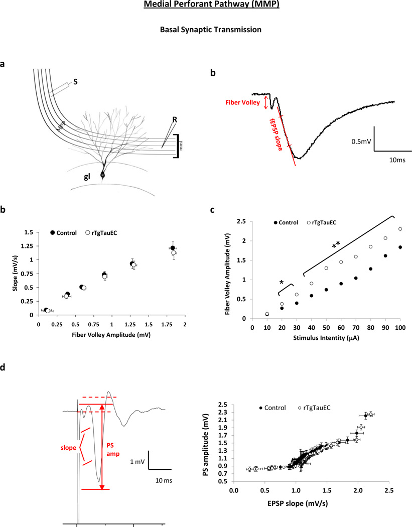
Fig. 4.
Synaptic transmission at the EC medial perforant pathway to dentate gyrus granule cell synapses (PP-DG). Extracellular field excitatory postsynaptic potentials (fEPSP) were recorded from horizontal EC-hippocampal slices of 16 month-old rTgTauEC and age-matched nontransgenic controls. a Schematic diagram of medial perforant pathway (MPP) synapto-architecture showing middle molecular layer (mml) and granule cells (gl) of the dentate gyrus with the positioning of stimulation (S) and recording (R) electrodes. b Typical shape of a fEPSP illustrating the method of assessment of presynaptic fiber volley amplitude and fEPSP slope. b Basal synaptic transmission was examined by building input-output (I/O) relationship at the PP-DG synapse in response to single electrical stimuli ranging from 10 to 800 A and plotted as the relationship between fEPSP slope and fiber volley amplitude, showing no significant difference between transgenic mice compared to control animals. c Fiber volley amplitude was plotted as a function of stimulus intensity and it shows a significant increase in fiber volley amplitude for a given stimulus intensity in the rTgTauEC mice compared to controls. (n= [control, n = 10 (3); rTgTauEC, n = 8 (4)]). d Typical shape of a population spike illustrating the method of assessment of amplitude of the population spike and the slope of the EPSP. E-S coupling was assessed by measuring the slope of the initial population EPSP and the amplitude of the population spike. Curves were generated for EPSP slope vs. population spike amplitude. There was no change in E-S coupling in rTgTauEC mice compared to controls the field suggesting that these animals do not have a post-synaptic change. (n= [control, n = 4 (3); rTgTauEC, n = 4 (3)]).

