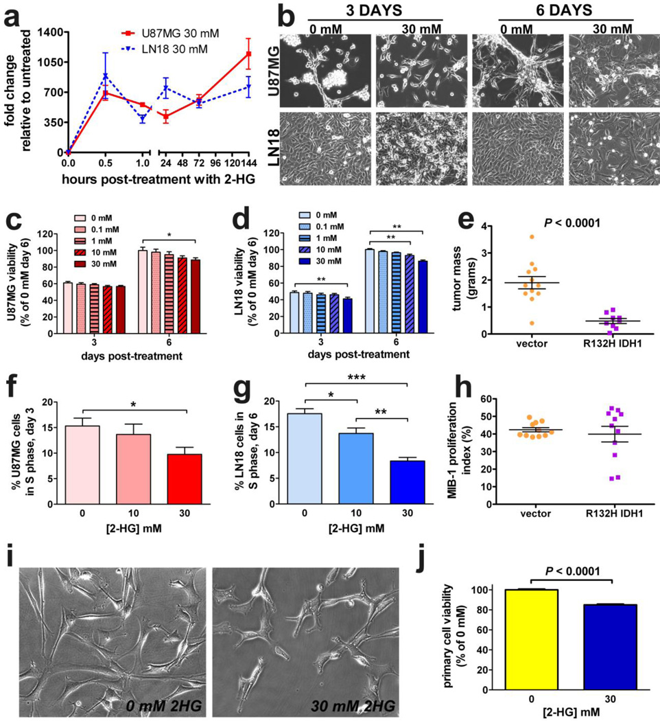
Fig. 1. 2-HG and mutant IDH1 inhibit glioma growth.
Both U87MG and LN18 cells were pulsed with 30 mM exogenous unmodified 2-HG in vitro. LC-MS of the cell lysates at various timepoints after treatment showed that 2-HG rapidly accumulated in cells and persisted through 6 days (a. Data represent mean ± SEM, n = 3. By phase contrast microscopy (b, both U87MG and LN18 cells showed signs of toxicity in response to 2-HG. Images are 200× magnification. Both U87MG (c) and LN18 cells (d) showed similar dose-dependent sensitivity to 2-HG as measured by MTS assay. For each cell type, data are normalized to the vehicle (0 mM) condition and are expressed as mean ± SEM, n=12. *P < 0.05; **P 0.001. (e) U87MG cells stably expressing R132H IDH1 were smaller than control tumors. Each dot represents a single xenograft, data are expressed as mean ± SEM, n = 11. In both U87MG (f) and LN18 cells (g), 2-HG suppressed entry into S-phase after 3 days of treatment. Data represent mean ± SEM, n = 5. *P < 0.05; **P < 0.01; ***P < 0.001. However, there was no significant difference in Mib-1 proliferation index between vector and R132H IDH1 xenografts (h), n = 11. Similar to the glioma cell lines, primary cultures of 2169 patient-derived GBM cells (IDH1 wild-type) were sensitive to 6 days of 2-HG as observed by phase contrast microscopy (i, 200×) and MTS assay (j).

