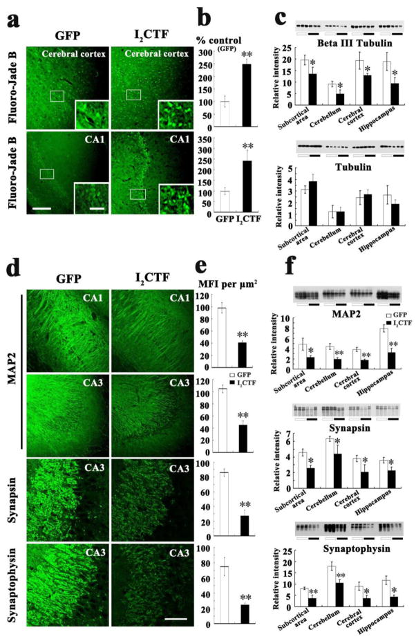
Fig. 7. Expression of I2CTF causes neurodegeneration and loss of neuronal plasticity.
a, b: Fluoro-Jade B staining showed an increase in neurodegeneration in I2CTF rats; c: Western blots and their quantitative analysis showed a decrease in the level of βIII tubulin and not in total tubulin in I2CTF rats; d, e: Immunohistofluorescent staining showed a decrease in the density of MAP2 in CA1 and CA3, synapsin 1 in CA3, and synaptophysin in CA3 of the hippocampi of I2CTF rats; and f: Western blots and their quantitative analysis showed a marked decrease in the levels of MAP2, synapsin 1, and synaptophysin in the I2CTF rats’ hippocampus, cerebral cortex, subcortical area (SA) and cerebellum. Data from 14-month-old rats. *p<0.05; **p<0.01. Scale bar: a: 300 μm; inset: 100 μm; d: 500 μm.

