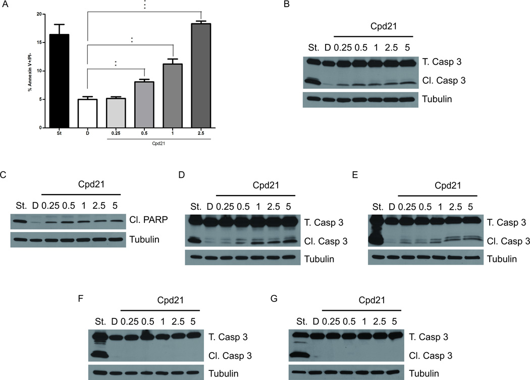
Figure 4. Cpd21 Induces Apoptosis in vitro.
(A) Annexin V staining as detected by flow cytometry. sMPNST cells were treated with DMSO, D, or Cpd21 (0.25, 0.5, 1, and 2.5 µM). sMPNST cells were treated with staurosporine, St, as positive control. (B–F) Western blotting for caspase 3 cleavage in sMPNST cells (B), PARP cleavage in sMPNST cells (C), caspase 3 cleavage in MPNSTs from cis-Nf1+/−;p53+/− mice (D), caspase 3 cleavage in human MPNST cell line, S462 (E), caspase 3 cleavage in Myc-SC (F), and caspase 3 cleavage in MEF cells (G). Each cell type was treated with staurosporine, St, as a positive control. Total caspase 3 (T. Casp 3) and cleaved caspase 3 (Cl. Casp 3) were assayed in each case. All values represent the mean±standard deviation. Student’s t-test used for significance testing (*p<0.05, **p<0.01, ***p<0.001).

