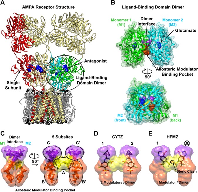
Figure 1.
Ionotropic glutamate receptor structure [PDB: 3KG2].6 (A) The ligand-binding domain (LBD) from 2 subunits (green and cyan) forms a dimer. (B) The LBD dimer [PDB: 1FTJ]4 has 2 agonist-binding sites (blue) and a symmetrical allosteric modulator-binding pocket that extends along the dimer interface. The accessible volume of the modulator-binding cavity is depicted by surfacing a composite of bound modulator structures (see Supporting Information). (C) The modulator-binding pocket can be divided into 5 subsites, A (yellow), B (orange), B′, C (purple), and C′. (D) Cyclothiazide (CYTZ) binds at two sites in the modulator-binding pocket with 2-fold symmetry. (E) Hydroflumethiazide (HFMZ) binding extends into the A subsite, obstructing the second binding site and allowing only 1 HFMZ to bind per dimer.

